The myotendinous junction (MTJ) is a complex specialized region located at the muscle-tendon interface that represents the primary site of force transmission. Despite their different embryologic origins, muscle and tendon morphogenesis occurs in close spatial and temporal association.
What is a Musculotendinous Junction?
The connection between a muscle and its tendon. The junction contains muscle cells with sarcomere membranes, which are folded inwards in a complicated manner.
What is the function of the Myotendinous Junction?
The myotendinous junction (MTJ) is the interface between muscle and tendon and where force is transmitted between the two tissues. It is also a common location for strain injuries in sports. Most of these can be prevented by heavy eccentric exercise.
What makes up the Musculotendinous Junction?
Each muscle has two tendons, one proximally and one distally. The point at which the tendon forms attachment to the muscle is also known as the musculotendinous junction (MTJ) and the point at which it attaches to the bone is known as the osteotendinous junction (OTJ).What is Musculotendinous structure?
The thoracoabdominal diaphragm is a composite musculotendinous structure, separating the thoracic and abdominal cavities. Reemphasis of the already well-delineated variations of the muscular and tendinous portions, and blood and nerve supply of the diaphragm is becoming apparent.
Where is the Achilles located on your body?
Overview and anatomy The Achilles tendon is a thick tendon located in the back of the leg. It connects the gastrocnemius and soleus muscles in the calf to an insertion point at the calcaneus (heel bone). It is the strongest tendon in the body and allows people to push off while walking, running and jumping.
What is the Musculotendinous unit?
Component. From a mechanical perspective, the musculotendinous unit behaves as a contractile component (muscle fibers) in parallel with one elastic component (muscle membranes) and in series with another elastic component (tendons).
Where does the vastus lateralis insertion?
Vastus lateralis The muscle descends through the lateral part of the anterior thigh compartment and inserts to the base of patella via the quadriceps tendon.What is the location of ligaments?
Ligaments are bands of tough elastic tissue around your joints. They connect bone to bone, give your joints support, and limit their movement. You have ligaments around your knees, ankles, elbows, shoulders, and other joints.
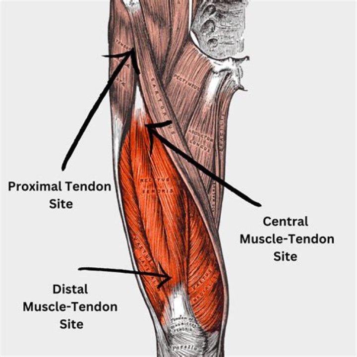
Where is rectus femoris?Rectus femoris is part of the quadriceps group. It is a bulk of muscle located in the superior, anterior middle compartment of the thigh and is the only muscle in the quadriceps group that crosses the hip.
Article first time published onHow Myotendinous junction is formed?
During embryogenesis, tendon cells attach to the developing muscle through the Extracellular Matrix (ECM) forming a specialized junction called Myotendinous Junction (MTJ) (Schweitzer et al., 2010; Subramanian and Schilling, 2015).
How do you get rid of tendinosis?
- Rest. …
- Adjust ergonomics and biomechanics. …
- Use appropriate support. …
- Stretch and keep moving, though conservatively. …
- Apply ice. …
- Eccentric strengthening. …
- Massage. …
- Nutrition.
What is a Myotendinous junction tear?
Purpose. Tears involving the myotendinous junction (MTJ) of the infraspinatus (IS) have been recently described on MRI. These occur centrally in the muscle belly, and are not associated with full thickness tears of the distal infraspinatus tendon.
What is Tendinous?
Definition of tendinous 1 : consisting of tendons : sinewy tendinous tissue. 2 : of, relating to, or resembling a tendon.
Which of the following detects the magnitude of mechanical stress at the Musculotendinous Junction?
The Golgi tendon organ detects the magnitude of mechanical stress at the musculotendinous junction.
What is Endomysium made of?
The endomysium, meaning within the muscle, is a wispy layer of areolar connective tissue that ensheaths each individual muscle fiber, or muscle cell. It also contains capillaries and nerves. It overlies the muscle fiber’s cell membrane: the sarcolemma.
Which of the following is the basic properties of the Musculotendinous unit?
Behavioral Properties of the Musculotendinous Unit The four behavioral properties of muscle tissue are extensibility, elasticity, irritability, and the ability to develop tension.
What is the ability to become short and thick?
Contractility. The ability to shorten and thicken (contract) when a stimulus is received.
Is the Achilles the ankle?
The Achilles tendon is the largest tendon in your body. It stretches from the bones of your heel to your calf muscles. You can feel it: a springy band of tissue at the back of your ankle and above your heel. It lets you point your toes toward the floor and raise up on your tiptoes.
How do you relieve Achilles tendon pain?
- Rest.
- Ice.
- Nonsteroidal anti-inflammatory drugs (NSAIDs) for pain relief, such as ibuprofen or naproxen.
- Specific exercises to strengthen your calf muscles.
- Physical therapy.
- Eccentric strength training. …
- Low-impact activities, such as swimming.
Why does Achilles heel hurt?
Most likely, your Achilles pain is caused by overuse of the tendon. It’s common that people who are athletic and active get Achilles pain the most. It’s not something that you should let go and keep doing activities on. This can make it worse and cause further issues and pain in your tendon.
Where is ligament in knee?
The ligaments in the knee connect the femur (thighbone) to the tibia (shin bone), and include the following: Anterior cruciate ligament (ACL). The ligament, located in the center of the knee, that controls rotation and forward movement of the tibia (shin bone). Posterior cruciate ligament (PCL).
Where is cartilage located?
Cartilage is a connective tissue found in many areas of the body including: Joints between bones e.g. the elbows, knees and ankles. Ends of the ribs. Between the vertebrae in the spine.
What is called ligament?
A ligament is the fibrous connective tissue that connects bones to other bones. It is also known as articular ligament, articular larua, fibrous ligament, or true ligament. Other ligaments in the body include the: Peritoneal ligament: a fold of peritoneum or other membranes.
Where is the vastus lateralis muscle in the thigh?
The vastus lateralis muscle is located on the lateral side of the thigh. This muscle is the largest of the quadriceps which includes: rectus femoris, vastus intermedius, and vastus medialis. Together, the quadriceps act on the knee and hip to promote movement as well as strength and stability.
Where is the middle third of the vastus lateralis muscle of the thigh?
The middle third of the lateral thigh between the trochanter major and the knee is an injection site in the vastus lateralis and is not in close proximity to any major blood vessels or nerves.
Where are quadriceps muscles located?
quadriceps femoris muscle, large fleshy muscle group covering the front and sides of the thigh. It has four parts: rectus femoris, vastus lateralis, vastus medialis, and vastus intermedius.
Is rectus femoris an organ?
A collective name of the four-headed skeletal muscle of the thigh, comprised of the rectus femoris, vastus intermedius, vastus lateralis, and vastus medialis. (quadriceps femoris) Organ cluster of muscle organs in the anterior compartment of thigh.
What is the rectus femoris for?
The rectus femoris is a biarticulate muscle, meaning it passes over two joints: the knee and hip. Its main function is as a knee extender; however, the proximal attachment at the anterior inferior iliac spine and the acetabulum allows for this muscle to act as a hip flexor as well.
What exercise works the rectus femoris?
A simple way to strengthen rectus femoris is to do slow straight leg raises. Simply lay on your back, bend one knee and place that foot on the floor, keep the other leg straight and very slowly lift the leg until both knees meet, hold for a few seconds then slowly lower your leg.
What is supraspinatus muscle?
Description. Supraspinatus is the smallest of the 4 muscles which comprise the Rotator Cuff of the shoulder joint specifically in the supraspinatus fossa. It travels underneath the acromion.
