It originates on the anterior surface of the manubrium
What bone does the Sternocleidomastoid attach to?
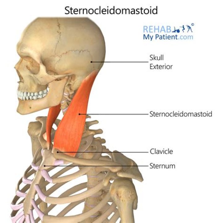
The sternocleidomastoid muscle is a two-headed neck muscle, which true to its name bears attachments to the manubrium of sternum (sterno-), the clavicle (-cleido-), and the mastoid process of the temporal bone (-mastoid).
Does the Sternocleidomastoid attach to the mandible?
Sternocleidomastoid: A thick rectangular muscle that is responsible for many movements within the neck. Attachments: Dual-headed, the sternocleidomastoid originates from the clavicle and the sternum and attaches to the mandible.
Where does the Sternocleidomastoid attach on sternum?
One attaches on the front (i.e., the anterior surface) of the manubrium. The manubrium is uppermost section of the breastbone. The other head attaches on the top part (called superior aspect) of the collarbone, near the midline of the body.Where does the Sternocleidomastoid insert quizlet?
Where does the sternocleidomastoid muscle originate and insert? ORIGINATES from the external surface of the occipital bone and posterior midline of the cervical and thoracic regions. INSERTS on the lateral third of the clavicle and parts of the scapula.
Where does levator scapulae attach?
Insertion. The levator scapulae inserts on to the vertebral margin of the scapula between the superior angle and the root of the spine.
Where does the sternocleidomastoid muscle attach superiorly?
Inferiorly, the muscle has two heads, tendinous sternal head attached to manubrium sterni and muscular clavicular head attached to medial third of the clavicle (clavicular head). Superiorly, muscle is attached to the mastoid process of the temporal bone and superior nuchal line of the occipital bone.
What muscles attach to the mandible?
It is the moving part of the jaws when the body is engaged in the feeding process and for that reason all the muscles of mastication including the medial and lateral pterygoid muscles, the temporal muscle and the masseter muscle attach to it. The mandible is only one of the skull bones.Where is the Platysma muscle located?
Dissimilar to other muscles of the body that lie deep to the subcutaneous tissue, the platysma is located within the subcutaneous tissue of the neck (superficial layer of the cervical fascia). Its superficial position means that surgical dissections of the neck must acknowledge the underlying neurovascular structures.
What muscles are connected to the jaw?The jaw muscles move the jaw in a complex three-dimensional manner during jaw movements. There are three jaw-closing muscles (masseter, temporalis, and medial pterygoid) and two jaw-opening muscles (lateral pterygoid and digastric). The basic functional unit of muscle is the motor unit.
Article first time published onWhat is the origin insertion and action of the sternocleidomastoid?
Structure. The sternocleidomastoid muscle originates from two locations: the manubrium of the sternum and the clavicle. It travels obliquely across the side of the neck and inserts at the mastoid process of the temporal bone of the skull by a thin aponeurosis.
What is the insertion of the pectoralis major quizlet?
Origin: Ribs 3-5 near costal cartilages, Inserts medial border and superior surface of coracoid process of scapula, innervation Medial Pectoral nerve, Fx, Stabilizes scapula by drawing it inferiorly and anteriorly against thoracic wall.
Which of the following is the insertion of the pectoralis major?
OriginClavicular part: anterior surface of medial half of clavicle Sternocostal part: anterior surface of sternum, Costal cartilages of ribs 1-6 Abdominal part: Anterior layer of rectus sheathInsertionCrest of greater tubercle of humerus
Which of the following describes muscle insertion?
Muscle insertion refers to a muscle’s distal attachment—the end of the muscle furthest away from the torso. For example, the bicep insertion occurs at the elbow.
What is the Sternohyoid?
The sternohyoid is a strap like infrahyoid muscle that connects the hyoid bone with the clavicle and sternum. … The function of this muscle is to reestablish the breathing process by pulling the hyoid bone and larynx inferiorly after deglutition. This article will discuss the anatomy of the sternohyoid muscle.
Where does the orbicularis oculi attach laterally?
From the orbital margin, the muscle extends inward and inserts on the lateral palpebral raphe, which is a ligament that is located on the outer part of the eye socket. The orbicularis oculi muscle inserts onto the lateral palpebral raphe which is located on the outer part of each eye socket.
Where does the soleus originate and insert?
OriginSoleal line, medial border of tibia, head of fibula, posterior border of fibulaInsertionPosterior surface of calcaneus (via calcaneal tendon)InnervationTibial nerve (S1, S2)VascularizationPosterior tibial artery and veinFunctionTalocrural joint: Foot plantar flexion
What is the distal attachment of levator scapulae?
The levator scapulae originates from the posterior tubercle of the transverse process of cervical vertebrae one to four. The muscle is inserted into medial border of the scapula extending from superior angle to junction of spine and medial border of scapula.
Where is the rhomboid minor?
Origin & Insertion The rhomboid minor is a cylindrical muscle that originates at the ligamentum nuchae and C7 and T1 vertebra. It inserts at the scapula’s medial border near the base of the spine of the scapula.
What is the insertion of the trapezius muscle?
Insertion. The muscle inserts on the lateral third of clavicle, acromion, and spine of scapula.
What is the sternocleidomastoid?
Sternocleidomastoid (SCM) (synonym musculus sternocleidomastoideus) is a paired superficial muscle in the anterior portion of the neck. The sternocleidomastoid muscle (SCM) is an important landmark in the neck which divides it into an anterior and a posterior triangle.
What is the inferior attachment insertion of the temporalis?
What is the inferior attachment/insertion of the temporalis? Temporal Fossa/Coronoid process and ramus of mandible.
Which of the following bones is not connected to the sternocleidomastoid?
What is the only extrinsic eye muscle to originate from the anterior orbit? … Which of the following bones is NOT attached to the sternocleidomastoid muscle? Mandible. What is the largest and most powerful muscle of the erector spinae muscle group?
Where is the Triangularis?
Muscles of the head, face, and neck (labeled as triangularis near chin). The depressor anguli oris muscle (triangularis muscle) is a facial muscle. It originates from the mandible and inserts into the angle of the mouth. It is associated with frowning, as it depresses the corner of the mouth.
What does the Platysma muscle connect to?
The platysma muscle is a muscle that begins at the upper chest/shoulder areas and extends upward through the sides of the neck and attaches to the skin around the mouth and mandible. In the neck, this muscle covers the sternocleidomastoid, which is a deep muscle that runs up vertically on each side of the neck.
Is the platysma and Sternocleidomastoid more anterior?
Platysma muscleArterybranches of the submental artery and suprascapular arteryNervecervical branch of the facial nerve
Which part of the mandible attaches to the cranium?
The mandible articulates with the cranium via the temporomandibular joint.
Is attached to the bones via tendons?
Skeletal muscles are attached to the skeleton by tough connective tissues called tendons(see Figure above). Many skeletal muscles are attached to the ends of bones that meet at a joint. The muscles span the joint and connect the bones. When the muscles contract, they pull on the bones, causing them to move.
What is the strongest muscle in the human body?
The strongest muscle based on its weight is the masseter. With all muscles of the jaw working together it can close the teeth with a force as great as 55 pounds (25 kilograms) on the incisors or 200 pounds (90.7 kilograms) on the molars.
Are jaw muscles connected to the neck?
Because muscles in your neck are connected to your jaw, muscle tension that starts in your TMJ can move to your neck. This causes aches, spasms, tension and reduced flexibility in your neck.
What is the muscle that inserts on the hyoid bone?
Mylohyoid muscle They interdigitate in the midline raphe anteriorly and posteriorly, they insert into the anterior part of the body of the hyoid bone.
