For example, sensory neurons send information from the eyes, ears, nose, tongue, and skin to the brain. Motor neurons carry messages away from the brain to the rest of the body.
What carries the message from the eye?
The retina then sends nerve signals are sent through the back of the eye to the optic nerve. The optic nerve carries these signals to the brain, which interprets them as visual images. The portion of the brain that processes visual input and interprets the messages that the eye sends is called the visual cortex.
What kind of neurons carry messages to the brain from your eyes and ears?
Sensory neurons carry information from the sense organs (such as the eyes and ears) to the brain.
Which type of neuron carries messages?
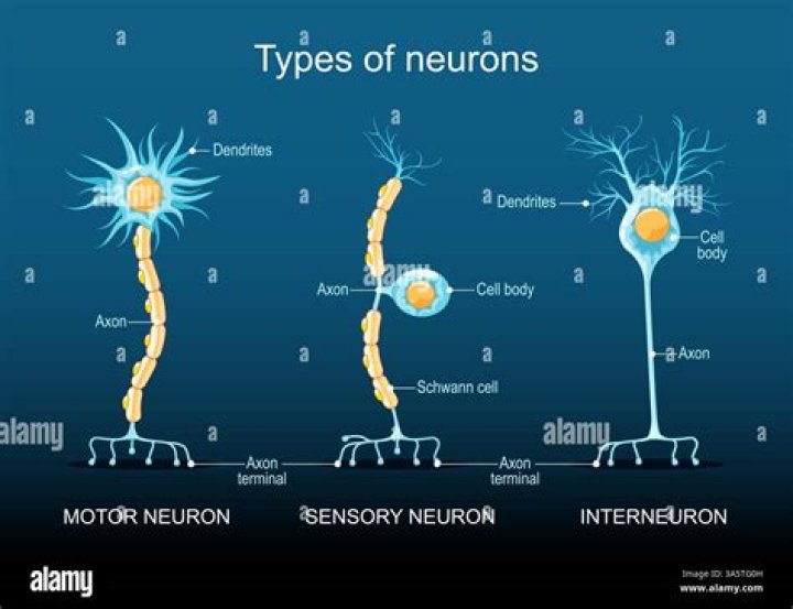
Three types of neurons occur. Sensory neurons typically have a long dendrite and short axon, and carry messages from sensory receptors to the central nervous system. Motor neurons have a long axon and short dendrites and transmit messages from the central nervous system to the muscles (or to glands).What nerve sends messages from the eye to the brain?
The optic nerve of each eye meets the other at the optic chiasm. Medial nerves of each optic nerve cross, but lateral nerves stay on the same side. The overlap of nerve fibers allows for depth perception. Electrical impulses are communicated to the visual cortex of the brain by way of the optic nerve.
What is sclera?
Listen to pronunciation. (SKLAYR-uh) The white layer of the eye that covers most of the outside of the eyeball.
What are retinas?
The retina contains millions of light-sensitive cells (rods and cones) and other nerve cells that receive and organize visual information. Your retina sends this information to your brain through your optic nerve, enabling you to see.
What part of the cerebrum processes messages from the eyes?
The occipital lobe, in the rear of the brain, processes light and other visual information from the eyes.How do neurons carry messages?
Your neurons carry messages in the form of electrical signals called nerve impulses. To create a nerve impulse, your neurons have to be excited. … So when a nerve impulse reaches the end of one neuron, a neurotransmitter chemical is released. It diffuses from this neuron across a junction and excites the next neuron.
Which nerve carries messages of touch from the tongue?Another cranial nerve (the trigeminal nerve, V) also innervates the tongue, but is not used for taste. Rather, the trigeminal nerve carries information related to touch, pressure, temperature and pain.
Article first time published onWhat is spiral cord?
The spinal cord is a long bundle of nerves and cells that extends from the lower portion of the brain to the lower back. It carries signals between the brain and the rest of the body. This article covers the key anatomy of the spinal cord and its functions. It also provides some information about spinal cord injuries.
What do glia cells do?
Primarily, glial cells provide support and protection to the neurons (nerve cells), maintain homeostasis, cleaning up debris, and forming myelin. They essentially work to care for the neurons and the environment they are in.
Which nerves carry messages to and from the brain?
Motor nerves carry messages from the brain and the spinal cord to other parts of the body.
Is it vagus or vagal nerve?
Vagus nerveTA26332FMA5731Anatomical terms of neuroanatomy
Which part of the neuron receives messages from other neurons?
Dendrites extend out from the cell body and receive messages from other nerve cells. An axon is a long single fiber that transmits messages from the cell body to the dendrites of other neurons or to other body tissues, such as muscles.
What is ciliary epithelium?
The ciliary body is a part of the eye that includes the ciliary muscle, which controls the shape of the lens, and the ciliary epithelium, which produces the aqueous humor. The aqueous humor is produced in the non-pigmented portion of the ciliary body.
What is macular thickening?
Macular edema is a swelling or thickening of the eye’s macula, the part of the eye responsible for detailed, central vision. The macula is a very small area at the center of the retina—a thin layer of light-sensitive tissue that lines the back of the eye.
What is retinal and opsin?
Retinal, bound to proteins called opsins, is the chemical basis of visual phototransduction, the light-detection stage of visual perception (vision). Some microorganisms use retinal to convert light into metabolic energy. … Retinal itself is considered a form of vitamin A when eaten by an animal.
What is the optic disc?
Optic disc: The circular area in the back of the inside of the eye where the optic nerve connects to the retina. Also called the optic nerve head.
What is blue sclera?
Blue sclera: a bluish coloration of the whites of the eyes. The blue color is caused by thinness and transparency of the collagen fibers of the sclera, allowing the veins in the underlying tissue to show through. Blue sclerae are characteristic of a number of conditions, particularly connective tissue disorders.
What causes Pinguecula?
Although a pinguecula itself is usually harmless, it sometimes causes redness or irritation to the eye. Chronic exposure to sun and ultraviolet radiation is thought to be the most common reason for the development of pinguecula, and it typically takes months or years.
How do neurons communicate quizlet?
Neurons communicate by sending messages using action potentials (electrically passing through their axons). Each neuron picks up signals at its dendrites, passes the signals down the aon, into the aon terminals, and into the synapses.
How does the neurons transmit information?
Neurons have a membrane featuring an axon and dendrites, specialized structures designed to transmit and receive information. Neurons release chemicals known as neurotransmitters into synapses, or the connections between cells, to communicate with other neurons.
Do neurons communicate electrically or chemically?
Neurons communicate with each other via electrical events called ‘action potentials’ and chemical neurotransmitters. At the junction between two neurons (synapse), an action potential causes neuron A to release a chemical neurotransmitter.
What does the frontal lobe do?
The frontal lobes are important for voluntary movement, expressive language and for managing higher level executive functions. Executive functions refer to a collection of cognitive skills including the capacity to plan, organise, initiate, self-monitor and control one’s responses in order to achieve a goal.
What does temporal lobe do?
The temporal lobes are also believed to play an important role in processing affect/emotions, language, and certain aspects of visual perception. The dominant temporal lobe, which is the left side in most people, is involved in understanding language and learning and remembering verbal information.
Which lobe is responsible for visual activity?
Each side of your brain contains four lobes. The frontal lobe is important for cognitive functions and control of voluntary movement or activity. The parietal lobe processes information about temperature, taste, touch and movement, while the occipital lobe is primarily responsible for vision.
Which cranial nerves carry taste information?
The facial nerve (CN VII) innervates the anterior two thirds of the tongue, the glossopharyngeal nerve (CN IX) innervates the posterior one third of the tongue, and the vagal nerve (CN X) carries taste information from the back part of the mouth, including the upper third of the esophagus.
Where is the ocular nerve?
The optic nerve begins at the optic disk, a structure that is 1.5 mm (0.06 inch) in diameter and is located at the back of the eye. The optic disk forms from the convergence of ganglion cell output fibres (called axons) as they pass out of the eye.
What is lingual nerve?
The lingual nerve is one of the sensory branches of the mandibular division of the trigeminal nerve. [5] It contains general somatic afferent nerve fibers and, after chorda tympani joins it, also carries general visceral efferent nerve fibers and special visceral afferent fibers.
What is posterior root ganglion?
A dorsal root ganglion (or spinal ganglion; also known as a posterior root ganglion) is a cluster of neurons (a ganglion) in a dorsal root of a spinal nerve. The cell bodies of sensory neurons known as first-order neurons are located in the dorsal root ganglia.
