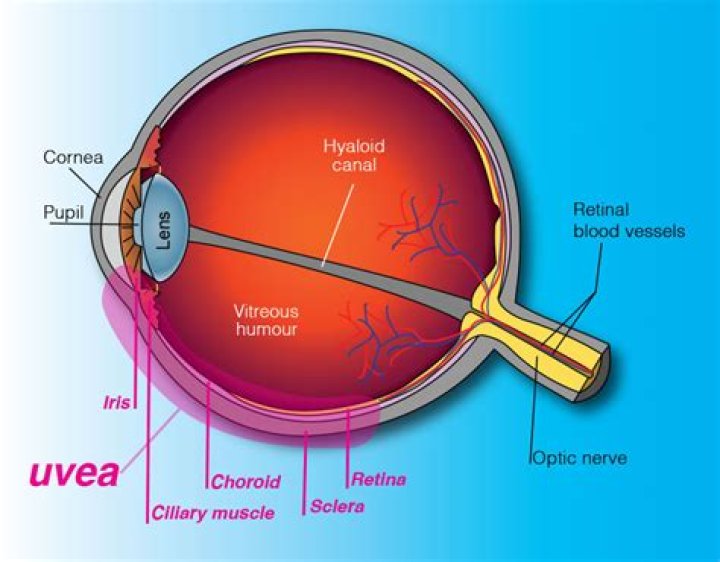
The uvea is the middle layer of tissue in the wall of the eye. It consists of the iris, the ciliary body and the choroid. When you look at your eye in the mirror, you will see the white part of the eye (sclera) and the colored part of the eye (iris). The iris is located inside the front of the eye.
Where is uvea in the eye?
The uvea consists of the layer and structures of the eye beneath the white of the eye (sclera).
Is retina part of uvea?
The uveal tract is the middle layer of the eye, divided into the anterior uvea (iris, ciliary body) and posterior uvea (choroid). The uvea is sandwiched between an outer layer (sclera) and an inner layer (retina). The anterior segment is separated from the posterior segment by the lens.
What is difference between choroid and uvea?
is that choroid is (anatomy) the vascular layer of the eye lying between the retina and the sclera while uvea is (anatomy) the middle of the three concentric layers that make up the eye; it is pigmented and vascular, and comprises the choroid, the ciliary body, and the iris.What is retina & uvea?
Home / Retina, Vitreous & Uvea. Retina (rɛtini from Latin rēte meaning “net”) is a light-sensitive layer at the back of the eye that covers about 65 percent of its interior surface. It is the third and inner coat of the eye which is a light-sensitive layer of tissue.
Where is uvea attached to sclera?
It has a rough outer surface which is attached to the sclera at the optic nerve and at the exit of the vortex veins. The smooth inner surface of the choroid is attached to the retinal pigmented epithelium (RPE). Choroid becomes continuous with pia and arachnoid at the optic nerve.
Is the uvea blue?
The middle coat of the eye is called the uvea (from the Latin for “grape”) because the eye looks like a reddish-blue grape when the outer coat has been dissected away.
Is the uvea a tissue?
uvea (or uveal tract), the middle layer of tissue surrounding the eye that consists of the iris, ciliary body, and choroid.What does uvea consist of?
The uvea is the middle layer of the eye. It lies beneath the white part of the eye (the sclera). It is made of the iris, ciliary body, and choroid. These structures control many eye functions, including adjusting to different levels of light or distances of objects.
What types of pathological processes of uvea do you know?Common pathologic changes involving the uvea include inflammatory and neoplastic diseases. Inflammatory changes are clinically recognized as various forms of uveitis. Among the neoplasms, both primary and metastatic tumors are found in all parts of the uvea.
Article first time published onWhat is the Colour of the uvea?
The originally medieval Latin term comes from the Latin word uva (“grape”) and is a reference to its grape-like appearance (reddish-blue or almost black colour, wrinkled appearance and grape-like size and shape when stripped intact from a cadaveric eye).
What is the posterior uvea?
Definition. Posterior uveitis is inflammation of the back part of the uvea known as the choroid. The uvea is the middle layer of the eye. Early treatment can improve outcomes. Normal Anatomy of the Eye.
Why is it called uveal tract?
The uveal tract, or simply uvea, is the pigmented middle membrane of the layers that make up the eye. The uveal tract is also called the vascular tunic of the eye because it is rich in its blood supply – i.e., vascular – and because it envelops the eye like a tunic would cover a body.
What is the surgery for a detached retina?
Vitrectomy This surgery is commonly used to fix a retinal detachment and is performed in an operating room. The vitreous gel, which is pulling on the retina, is removed from the eye and usually replaced with a gas bubble.
Do humans have photoreceptor cells?
There are currently three known types of photoreceptor cells in mammalian eyes: rods, cones, and intrinsically photosensitive retinal ganglion cells. … In humans, there are three different types of cone cell, distinguished by their pattern of response to light of different wavelengths.
What are EOG and ERG?
The electroretinogram (ERG) and electrooculogram (EOG) are electrophysiological tests employed in ophthalmology to diagnose degeneration or injury to the outer half of the retina, including the rods and cones of the visual system.
Is the choroid the uvea?
The uvea or vascular tunic of the eye consists of the choroid, ciliary body, and iris. The choroid lies between the sclera and RPE, and contains connective tissue, capillaries, and melanocytes. The choroid terminates anteriorly as the ciliary body.
What is a ciliary flush?
Ciliary flush is usually present in eyes with corneal inflammation, iridocyclitis or acute glaucoma, though not simple conjunctivitis. A ciliary flush is a ring of red or violet spreading out from around the cornea of the eye.
Where is ciliary muscle?
The ciliary muscle is elongated, triangular in shape, and located beneath the anterior sclera just posterior to the limbus. The shortest side of the triangular region faces anterior-inward and it is to this region of the ciliary body that the base of the iris inserts.
What produces aqueous humor?
Aqueous humour is made by the ciliary body. Strands from the ciliary body support the lens behind the coloured part of the eye (iris). Aqueous humour flows into the front of the eye through the pupil. Aqueous humour drains out of the eye through the trabecular meshwork.
What is the ciliary body?
A part of the middle layer of the wall of the eye. The ciliary body is found behind the iris and includes the ring-shaped muscle that changes the shape of the lens when the eye focuses. It also makes the clear fluid that fills the space between the cornea and the iris.
Is there a permanent cure for uveitis?
Treatment Strategies Even though there may not be a permanent cure for Uveitis, each attack can be treated, depending upon the cause, severity and location of the inflammation. The treatment may include eye drops, injections under the eye or oral medication.
What is granulomatous uveitis?
Granulomatous uveitis is an inflammation of the uveal tract characterized by the formation of granulomas due to infectious or non-infectious causes.
Is ciliary body transparent?
The ciliary body is a ring-shaped thickening of tissue inside the eye that divides the posterior chamber from the vitreous body. … The inner layer is transparent and covers the vitreous body, and is continuous from the neural tissue of the retina.
What does posterior uveitis feel like?
Symptoms that people may experience if they have posterior uveitis include: Floaters. Reduced visual acuity (sharpness of vision) Light sensitivity.
How long does it take to go blind from uveitis?
The mean duration of visual loss was 20.35 months for patients with moderate visual loss and 22.8 months in patients with severe loss of vision. In patients with unilateral visual loss the mean duration was 20 months whereas it was 42.61 months in patients with bilateral visual morbidity.
Does posterior uveitis go away?
The part of your eye affected by uveitis will determine the duration of the condition. With proper treatment, anterior uveitis can clear up in a matter of days to weeks. Posterior uveitis, on the other hand, may last several months or years and could permanently alter your vision.
What are retinas?
The retina is a layer of tissue in the back of your eye that senses light and sends images to your brain. In the center of this nerve tissue is the macula. It provides the sharp, central vision needed for reading, driving and seeing fine detail. Retinal disorders affect this vital tissue.
What is pars plicata?
The pars plicata is the portion of the ciliary body that is responsible for producing aqueous humor, the fluid of the anterior chamber. The production of too much aqueous humor, or reabsorption that occurs too slowly, can lead to increases in the pressure within the eye.
How long does it take to recover from detached retina surgery?
You will need 2 to 4 weeks to recover before returning to your normal activities. This care sheet gives you a general idea about how long it will take for you to recover. But each person recovers at a different pace.
What are the warning signs of a detached retina?
- Dots or lines (floaters) suddenly appear in your vision or suddenly increase in number.
- Flashes of light in your vision.
- Dark ‘curtain’ or shadow moving across your vision.
