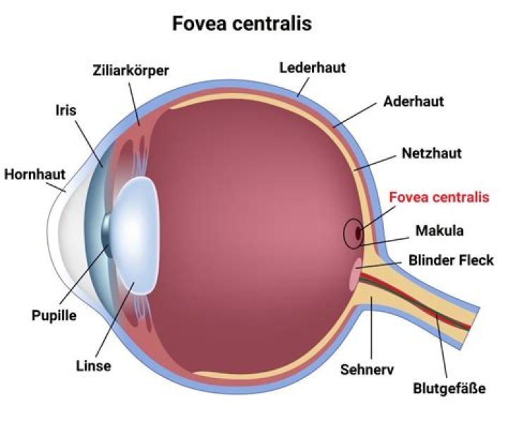
The fovea centralis is located in the center of the macula lutea
What is the structure and function of the fovea?
A fovea is a pitted invagination in the inner retina (fovea interna) that overlies an area of photoreceptors specialized for high acuity vision (fovea externa). A fovea contains particularly high numbers of photoreceptors and neurons, and provides the highest visual resolution (Walls, 1942; Polyak, 1957).
Is the fovea centralis the blind spot?
The blind spot (Fovea centralis) The blind spot, or scotoma, is the place in our eyes where the optic nerve passes through the retina to the brain. … The blind spot is located about 15 degrees on the nasal side of the fovea.
What does the fovea centralis contain?
In the center of the macula is the fovea centralis. The macula contains mostly cones and few rods, and the fovea centralis contains only cones and no rods.How is the fovea centralis different from the rest of the retina?
It has four anatomical characteristics which distinguish it from the surrounding retina and are modifications to support high acuity: … The foveal center or ‘foveola’ contains the highest density of cone photoreceptors in the retina. Cone photoreceptors function in bright light and support high acuity and color vision.
What is the difference between macula and fovea?
The macula is the pigmented part of the retina located in the very center of the retina. In the center of the macula is the fovea, perhaps the most important part of the eye. The fovea is the area of best visual acuity. It contains a large amount of cones—nerve cells that are photoreceptors with high acuity.
What is the function of the macula in the human eye?
The macula is part of the retina at the back of the eye. It is only about 5mm across but is responsible for our central vision, most of our colour vision and the fine detail of what we see. The macula has a very high concentration of photoreceptor cells – the cells that detect light.
What is the fovea centralis quizlet?
FOVEA CENTRALIS (MACULA LUTEA) portion of retina that where light is most focused when eye is looking directly at an object; the area of highest visual acuity; contains only cone cells. OPTIC DISC.How does the eye direct light onto the fovea centralis?
Photosensitive cells called rods and cones in the retina convert incident light energy into signals that are carried to the brain by the optic nerve. … In the middle of the retina is a small dimple called the fovea or fovea centralis.
What is the function of the blind spot?What is the purpose of a blind spot in the eye? The blind spot is where the optic nerve and blood vessels leave the eyeball. The optic nerve is connected to the brain. It carries images to the brain, where they’re processed.
Article first time published onWhat is the Foreva?
Fovea: In the eye, a tiny pit located in the macula of the retina that provides the clearest vision of all. Only in the fovea are the layers of the retina spread aside to let light fall directly on the cones, the cells that give the sharpest image. Also called the central fovea or fovea centralis.
Is incoming light focused on the optic disk of the fovea centralis?
When you stare directly at something, the image is focused on the fovea centralis part of the retina to maximize the visual acuity. The lens is not part of the retina, but rather it is in front of the retina. Its role is to focus incoming light beams onto the retina.
What is the difference between fovea and Foveola?
In context|anatomy|lang=en terms the difference between fovea and foveola. is that fovea is (anatomy) the retinal fovea, or fovea centralis, responsible for sharp central vision while foveola is (anatomy) the center of the fovea in the macula of the eye, approximately 035 mm in diameter, containing only cone cells.
What is different about the fovea and the rest of the retina why is visual acuity best when images fall on the fovea?
Relatively few photoreceptors feed each ganglion cell in the fovea, resulting in a low ratio, which maximizes visual acuity. … This allows light to strike the photoreceptors without passing through the other layers of retinal cells, minimizing light scatter that can blur the image.
Is the fovea responsible for central vision?
The fovea is responsible for sharp central vision (also called foveal vision), which is necessary in humans for activities for which visual detail is of primary importance, such as reading and driving.
What is macula Cirrus?
The Zeiss Cirrus HD-OCT is a non-invasive technology used for imaging the vitreous and retina — the multi-layered sensory tissue lining the back of the eye. The Optical Coherence Tomography (OCT) scanner provides physicians with an automated, segmented representation of the choroid and retinal layers.
Where is the macula in the fundus?
A fundus photo, showing the optic disc as a bright area on the right where blood vessels converge. The spot to the left of the centre is the macula.
What is fovea psychology?
a small depression in the central portion of the retina in which retinal cone cells are most concentrated and an image is focused most clearly. Also called fovea.
What is the difference between the fovea and the blind spot?
Visual acuity such as sharpness and detail is greatest at the fovea, while at the blind spot it is insensitive to visual stimulation, it’s the part of the retina that converges to the optic nerve.
Why is vision sharpest in the fovea?
The fovea is a highly specialized region of the retina. It is the spot of highest visual acuity in the eye and produces the sharpest vision and greatest color discrimination. The resolution or sharpness in vision is because of the high concentration of cone cells in the fovea.
What causes the Choroids dark color?
Melanin, a dark colored pigment, helps the choroid limit uncontrolled reflection within the eye that would potentially result in the perception of confusing images. In humans and most other primates, melanin occurs throughout the choroid.
What happens when light falls on the retina?
When light falls on the retina after being inverted by the lens, the incident light energy is converted by cells called rods and cones into electro-chemical signals. … These electro-chemical signals are then passed via the optic nerve to the brain which then interprets the signals to enable sight.
How do eyes perceive and interpret the rays of light in order to form an image?
When light hits the retina (a light-sensitive layer of tissue at the back of the eye), special cells called photoreceptors turn the light into electrical signals. These electrical signals travel from the retina through the optic nerve to the brain. Then the brain turns the signals into the images you see.
What is the sclera's function quizlet?
what is the sclera’s function? it maintains the shape and rigidity of the eyeball and attaches to the extrinsic eye muscles.
What is the function of the fovea quizlet?
-The fovea is responsible for sharp central vision (also called foveal vision), necessary for activities where visual detail is of primary importance, such as reading and driving. photoreceptors. -location where ganglion cell axons exit the eye to form the optic nerve.
Which of these are the functions of the ciliary body quizlet?
- ciliary body. has three functions: accommodation, aqueous humor production and the resorption, and maintenance of the lens zonules. …
- ciliary muscle. adjusts the shape of the lenses in order to focus the eyes.
- cornea. …
- pupil. …
- conjunctiva. …
- aqueous chamber. …
- iris. …
- lens.
What is the function of blind spot Class 10?
blind spot, small portion of the visual field of each eye that corresponds to the position of the optic disk (also known as the optic nerve head) within the retina. There are no photoreceptors (i.e., rods or cones) in the optic disk, and, therefore, there is no image detection in this area.
What is the blind spot and why is it so called?
Your retina is covered in light-sensitive cells, which send messages to your brain about what you see. Everyone has a spot in the retina where the optic nerve connects. In this area, there are no light sensitive cells, so this part of your retina cannot see. We call this the blind spot.
What is the blind spot in the eye and how does it impact the transduction of light energy?
The eye’s retina receives and reacts to incoming light and sends signals to the brain, allowing you to see. One part of the retina, however, doesn’t give you visual information—this is your eye’s “blind spot.”
What is the function of the optic nerve?
optic nerve, second cranial nerve, which carries sensory nerve impulses from the more than one million ganglion cells of the retina toward the visual centres in the brain. The vast majority of optic nerve fibres convey information regarding central vision.
What is the function of the optic disc?
The optic disc identifies the start of the optic nerve where messages from cone and rod cells leave the eye via nerve fibres to the optic centre of the brain. This area is also known as the ‘blind spot’. Optic nerve: leaves the eye at the optic disc and transfers all the visual information to the brain.
