Two bundle branches carry the electrical signal to the bottom of the heart and cause the ventricles to beat. These are termed the right bundle and left bundle.
What is the function of the bundle branches quizlet?
What is the primary function of the bundle branches? To conduct electrical activity from the Bundle of His down to the Purkinje network.
What is the role of the bundle of His bundle branches and Purkinje fibers?
The bundle of His has two branches: Left bundle branch sends electrical signals through the Purkinje fibers to your left ventricle. Right bundle branch sends electrical signals through the Purkinje fibers to your right ventricle.
What do the bundle branches in the septum do?
The bundle of His consists of wide, fast-conducting muscle fibers that carry the cardiac impulse through the insulating annulus fibrosus into the fibrous upper part of the ventricular septum. … The right bundle branch travels along the right side of the septum and supplies excitation to the right ventricle.What is the function of Purkinje fibers quizlet?
What is the function of the purkinje fibers? Send nerve impulses to the cells in the ventricles of the heart and cause them contract and pump blood either to the lungs or the rest of the body.
What is the purpose of the bundle of His in the heart conduction system?
The atrioventricular bundle (bundle of His) is a continuation of the specialised tissue of the AV node, and serves to transmit the electrical impulse from the AV node to the Purkinje fibres of the ventricles.
In what region of the heart are bundle branches located?
The bundle branches, or Tawara branches, are offshoots of the bundle of His in the heart’s ventricle. They play an integral role in the electrical conduction system of the heart by transmitting cardiac action potentials from the bundle of His to the Purkinje fibers.
What is the bundle branch made of?
A bundle branch block is either a complete or a partial interruption of the electrical pathways inside the wall of the heart between the two lower chambers (ventricles). The master pacemaker of the heart is the sinoatrial node, a small mass of muscle cells at the top of the right chamber (atrium) of the heart.What is bundle branch block ECG?
Bundle branch block is a condition in which there’s a delay or blockage along the pathway that electrical impulses travel to make your heart beat.
What is the function of Purkinje fibers in the heart?Purkinje fibers play a major role in electrical conduction and propagation of impulse to the ventricular muscle.
Article first time published onWhat is the meaning of bundle of His?
hĭs. The bundle of cardiac muscle fibers that conducts the electrical impulses that regulate the heartbeat, from the atrioventricular node in the right atrium to the septum between the ventricles and then to the left and right ventricles.
Where are the bundle branches located quizlet?
There are two branches of the bundle of His: the left bundle branch and the right bundle branch, both of which are located along the interventricular septum. The left bundle branch further divides into the left anterior fascicles and the left posterior fascicles.
What are the functions of the AV node?
The AV node controls the passage of the heart’s electrical signal from the atria to the ventricles. After an electrical impulse is generated by the sinus node (located at the top of the right atrium), it spreads across both atria, causing these chambers to beat.
What is the role of the Purkinje system in causing the ventricular muscle to synchronously contract?
The His-Purkinje System (HPS) is responsible for the rapid electric conduction in the ventricles. It relays electrical impulses from the atrioventricular node to the muscle cells and, thus, coordinates the contraction of ventricles in order to ensure proper cardiac pump function.
What system in the body do the SA node AV node bundle of His and Purkinje fibers belong to?
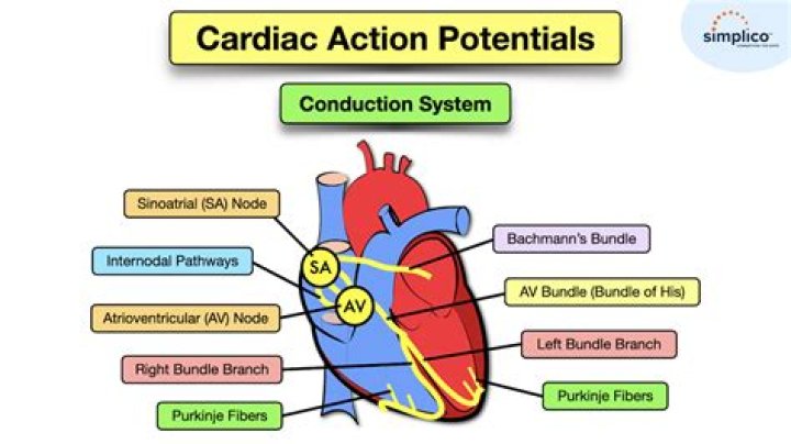
This group of muscle cells is called the cardiac conduction system. The main parts of the system are the SA node, AV node, bundle of HIS, bundle branches, and Purkinje fibers.
What is the difference between bundle of His and Purkinje Fibres?
The key difference between Bundle of His and Purkinje Fibres is that Bundle of His is a collection of specialized heart muscle cells for electrical conduction while Purkinje fibres are thin filaments originating from the left posterior fascicle and left anterior fascicles that help in distributing impulses to the …
What is the function of the heart in the cardiovascular system?
The heart is a pump, usually beating about 60 to 100 times per minute. With each heartbeat, the heart sends blood throughout our bodies, carrying oxygen to every cell. After delivering the oxygen, the blood returns to the heart. The heart then sends the blood to the lungs to pick up more oxygen.
Can right bundle branch block cause chest pain?
RBBB is usually an incidental finding on an ECG, which would have been carried out for another reason. However, in the presence of symptoms like chest pain or shortness of breath or syncope, it might signify underlying heart or lung disorders such as: Long standing right heart failure.
What does a right bundle branch block indicate?
Right bundle branch block (RBBB) is an abnormal pattern that is seen on the electrocardiogram (ECG), which indicates that the heart’s electrical impulse is not being distributed normally across the ventricles.
Can high blood pressure cause right bundle branch block?
Right bundle branch block can result from a number of conditions, such as: Heart disease due to high blood pressure in the lungs (pulmonary hypertension) Chronic obstructive lung disease (COPD) Blood clot in the lung (pulmonary embolism)
How many bundle branches does the heart have?
The bundle of His divides into two bundle branches. The left bundle branch conducts impulses to the left ventricle, and the right bundle branch conducts impulses to the right ventricle.
Are bundle branches nerves?
Bundle of HisTA23955FMA9484Anatomical terminology
Which artery is the main supplier for the bundle branches?
The atrioventricular bundle branch arose from the right coronary artery in 10% of cases, the left coronary artery in 73%, and both coronary arteries in 17%.
What is the function of Purkinje cells?
Purkinje cells participate in the processes of motor control and learning. They are the only cells that emit signals from the cerebellar cortex that is the outer layer of the cerebellum, though they can receive input from hundreds of thousands of cells.
What is the difference between Purkinje cells and Purkinje fibers?
The purkinje fibres are found in the sub-endocardium. They are larger than cardiac muscle cells, but have fewer myofibrils, lots of glycogen and mitochondria, and no T-tubules. These cells are connected together by desmosomes and gap junctions, but not by intercalated discs. Take a look at this EM of a purkinje cell.
What is Purkinje Fibres tissue?
Purkinje fibers or Purkinje cardiomyocytes are part of the whole complex of the cardiac conduction system, which is today classified as specific heart muscle tissue responsible for the generation of the heart impulses.
Where is bundle of His present?
The bundle of his is a network of the muscle fibers found in the wall of ventricles .
What is the moderator band?
In the human heart, the moderator band, or trabecula septomarginalis, is a muscle column that courses inferiorly from the right portion of the interventricular septum to the base of the anterior papillary muscle of the right ventricle This muscular structure is crossed by one or more arteries, which come from the …
What carries the wave of depolarization from the atrioventricular node to the bundle branches?
The conduction system of the heart. Left: Normal excitation originates in the sinoatrial (SA) node then propagates through both atria. The atrial depolarization spreads to the atrioventricular (AV) node, and passes through the bundle of His to the bundle branches/Purkinje fibers.
How are impulses transmitted through the heart quizlet?
In the normal heart, electrical impulses are initiated in the sinoatrial node (SA node), conducted through both atria, and directed to the atrioventricular node (AV node). The AV node delays transmission of the signal to the ventricles, allowing them to completely fill with blood.
What is the area of conduction in the heart quizlet?
Located between atria and ventricles superior to the interventricular septum; reeives impulses from AV node and conducts them to the bundle branches.
