The renal sinus is a cavity within the kidney which is occupied by the renal pelvis, renal calyces, blood vessels, nerves and fat • The renal hilum is the entry and exit site for structures servicing the kidneys: vessels, nerves, lymphatics, and ureters.
Is renal sinus the hilum?
The renal sinus is a fatty compartment located within the medial aspect of the kidney. It communicates with the perinephric space. It contains the renal hilum and is bordered by renal parenchyma laterally.
What is an alternate name for renal hilum?
Kidney anatomy, with hilum labeled at upper left. The renal hilum (Latin: hilum renale) or renal pedicle is the hilum of the kidney, that is, its recessed central fissure where its vessels, nerves and ureter pass.
What is the renal sinus also known as?
…the kidney known as the renal (kidney) sinus. The hilus is the point of entry and exit of the renal arteries and veins, lymphatic vessels, nerves, and the enlarged upper extension of the ureters.What is the main function of renal sinus?
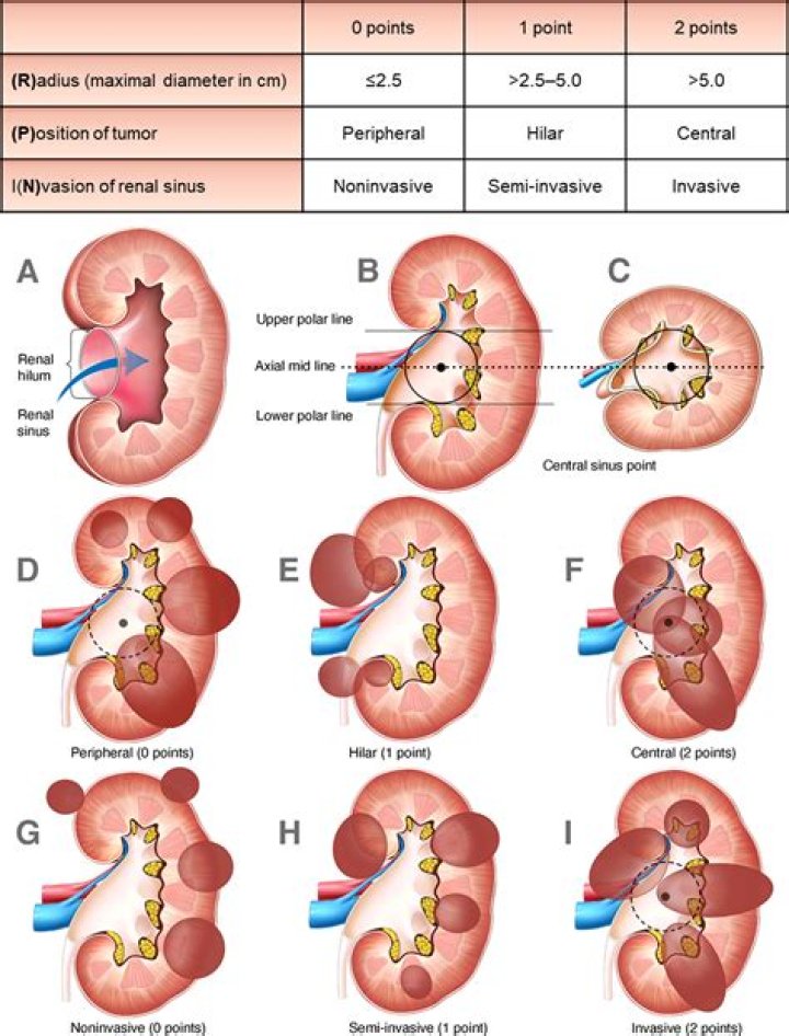
Fatty compartment along the hilus of kidney that invests the renal calyces, pelvis, blood vessels, lymphatics and nerves (image A) & (image B). Renal sinus abuts the renal cortical columns of Bertin without a connective tissue interface. Important landmark for staging renal carcinoma (extrarenal extension).
What is the function of the renal hilum?
The hilum is the concave part of the bean-shape where blood vessels and nerves enter and exit the kidney; it is also the point of exit for the ureters—the urine-bearing tubes that exit the kidney and empty into the urinary bladder. The renal pelvis connects the kidney to the rest of the body.
Is the renal papilla part of the renal sinus?
structure of human kidney sinus is known as a renal papilla. The bases of these pyramids are irregular, with slender striations extending toward the external kidney surface. The paler, more granular tissue external to the medulla is the cortex.
What is perirenal fat and what is its function?
What is the purpose of the perirenal fat capsule? It’s the fatty cushion that prevents kidney from being damaged or even rupturing due to trauma/injuries.What surrounds the renal sinus?
The hilum leads to a large cavity, called the renal sinus, within the kidney. The ureter and renal vein leave the kidney, and the renal artery enters the kidney at the hilum. The outer, reddish region, next to the capsule, is the renal cortex. This surrounds a darker reddish-brown region called the renal medulla.
What is the difference between renal pelvis and hilum?The renal hilum (Figure 2) is the entry and exit site for structures servicing the kidneys: vessels, nerves, lymphatics, and the ureters. … Emerging from the hilum is the renal pelvis, which is formed from the major and minor calyxes in the kidney.
Article first time published onAre renal columns part of the medulla?
The medulla consists of multiple pyramidal tissue masses, called the renal pyramids. In between the pyramids are spaces called renal columns through which the blood vessels pass.
Which of the following is correct about hilum of kidney?
Explanation: The hilum is on the most superior surface of the kidney. The hilum is where the ureter exits the kidney. The hilum is the concave indention of the kidney.
Where is renal pyramid?
Renal pyramids are kidney tissues that are shaped like cones. Another term for renal pyramids is malpighian pyramids. Between seven and eighteen pyramids exist in the innermost part of the kidney, which is called the renal medulla; in humans, there are usually only seven of the pyramids.
What is the difference between renal papillae and renal crest?
Macroscopically, the inner medulla can be subdivided into the base and the papilla or renal crest. The base is adjacent to the outer medulla. The papilla, or renal crest, is the terminal portion of the inner medulla, which extends into the renal pelvis or calices.
What is the difference between renal pelvis and renal papilla?
Renal PelvisRenal papillaRenal pelvis is the portion formed by the union of two or three calyces.It is the location where the renal pyramids present in the medulla empty urine into the minor calyx.
What is renal papilla in kidney?
The renal papillae are the areas where the openings of the collecting ducts enter the kidney and where urine flows into the ureters.
What are the two main function of renal tubule?
The major function of tubules is reabsorption and the process can either be through active transport or passive transport. In addition, secretions by tubules help in the urine formation without affecting the electrolyte balance of the body.
What is the difference between renal artery and renal vein?
The renal artery brings blood to the kidney for filtration whereas the renal vein carries away filtered blood from the kidney. The renal artery takes blood to the kidney. The renal vein takes blood away from the kidney.
What is the renal hilum and what three vessels help make up the renal hilum?
II Structure and Function of the Kidney The nerves, ureters, renal blood vessels, and the lymphatic vessels enter and leave the kidneys at their inner, concave, side that contains the renal hilum.
What are the three layers of kidney?
The Kidneys Are Composed of Three Main Sections Each kidney consists of an outer renal cortex, an inner renal medulla, and a renal pelvis.
What is renal sinus Lipomatosis?
Renal sinus lipomatosis is the accumulation of excessive nontumorous fatty tissue within the renal sinus. In the normal young adult, a thin layer of loose fatty tissue envelops the pelvocalyceal and vascular structures which traverse the sinus.
What is the importance of the perirenal fat capsule that surrounds the kidney?
Perinephric fat, also known as perirenal fat or the adipose capsule of the kidney, is a layer of fatty material that surrounds the kidneys. It plays an important role because it helps to cushion and protect the kidneys. Trace the pathway a creatinine molecule takes from a glomerulus to the urethra.
What is Perirenal fascia?
The perirenal fascia is a dense, elastic connective tissue sheath that envelops each kidney and adrenal gland together with a layer of surrounding perirenal fat forming the perirenal space.
Where is perirenal fat located?
Perirenal fat is located between the kidney capsule and the renal fascia. Both, perirenal adipose tissue and kidney cortex receive blood from the abdominal aorta.
Which structure separates the renal pyramids?
A frontal section through the kidney reveals an outer region called the renal cortex and an inner region called the renal medulla (Figure 25.1. 2). In the medulla, 5-8 renal pyramids are separated by connective tissue renal columns.
Why is the renal cortex darker than the medulla?
Cortex and medulla The renal cortex is the outer layer of the kidney tissue. It is darker than its underlying renal medulla because it receives over 90% of the kidney blood supply. The cortex has a grainy appearance, as it mostly contains ovoid and coiled parts of the nephrons (renal corpuscles and convoluted tubules).
Which of the following is incorrect about hilum?
Column IColumn IIB. Hilum(ii) Basal part of ovuleC. Integument(iii) One or two protective layers of ovuleD. Chalaza(iv) Region where body of ovule fuses with funicleE. Nucellus(v) Stalk of ovule
Which part of the nephron is involved in filtration?
Each nephron has a glomerulus, the site of blood filtration. The glomerulus is a network of capillaries surrounded by a cuplike structure, the glomerular capsule (or Bowman’s capsule).
Why are the kidneys referred to as Osmoregulatory structures?
Kidneys regulate the osmotic pressure of a mammal’s blood through extensive filtration and purification in a process known as osmoregulation. All the blood in the human body is filtered many times a day by the kidneys. … Kidneys eliminate wastes from the body; urine is the filtrate that exits the kidneys.
Are medulla and renal pyramid same?
The medulla is divided into 8-18 conical regions, called the renal pyramids; the base of each pyramid starts at the corticomedullary border, and the apex ends in the renal papilla which merges to form the renal pelvis and then on to form the ureter.
What is in renal pyramids?
The pyramids consist mainly of tubules that transport urine from the cortical, or outer, part of the kidney, where urine is produced, to the calyces, or cup-shaped cavities in which urine collects before it passes through the ureter to the bladder. The point of each pyramid, called the papilla, projects into a calyx.
