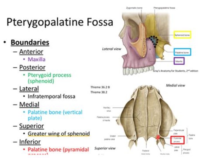
The pterygopalatine fossa (PPF) is a small, clinically inaccessible, fat-filled space located in the deep face that serves as a major neurovascular crossroad between the oral cavity, nasal cavity, nasopharynx, orbit, masticator space, and the middle cranial fossa.
What is present Pterygoid fossa?
The lateral and medial pteryoid plates (of the pterygoid process of the sphenoid bone) diverge behind and enclose between them a V-shaped fossa, the pterygoid fossa. This fossa faces posteriorly, and contains the medial pterygoid muscle and the tensor veli palatini muscle.
Why is the pterygopalatine fossa important?
The pterygopalatine fossa is an important pathway for the spread of neoplastic and infectious processes: medially: communicates with the nasal cavity via the sphenopalatine foramen, which transmits the sphenopalatine artery, the nasopalatine nerve and the posterior superior nasal nerves.
What passes through Pterygoid fossa?
Pterygoid and Pharyngeal Canals These two canals, along with the foramen rotundum, are the three openings in the posterior wall of the pterygopalatine fossa: Pterygoid canal – runs from the middle cranial fossa and through the medial pterygoid plate. It carries the nerve, artery and vein of the pterygoid canal.Is Pterygoid fossa same as pterygopalatine fossa?
Pterygopalatine fossaLatinfossa pterygopalatinaMeSHD056739TA98A02.1.00.025TA2429
What is scaphoid fossa?
Medical Definition of scaphoid fossa : a shallow oval depression that is situated above the pterygoid fossa on the pterygoid process of the sphenoid bone and that provides attachment for the origin of the tensor veli palatini muscle.
What is meant by fossa?
: an anatomical pit, groove, or depression the temporal fossa of the skull the fossa in the liver for the gallbladder.
Where is pterygoid fossa located?
The pterygopalatine fossa (PPF) is a small, clinically inaccessible, fat-filled space located in the deep face that serves as a major neurovascular crossroad between the oral cavity, nasal cavity, nasopharynx, orbit, masticator space, and the middle cranial fossa.What does Pterygoid Canal communicate?
The pterygoid canal is located on the posterior wall of the pterygopalatine fossa, between the foramen rotundum and the palatine canal. It communicates with the middle cranial fossa and from there, it transmits the nerve, artery, and vein of the pterygoid canal.
What is the palatine canal?The greater palatine canal (or pterygopalatine canal) is a passage in the skull that transmits the descending palatine artery, vein, and greater and lesser palatine nerves between the pterygopalatine fossa and the oral cavity.
Article first time published onWhat does pterygopalatine mean?
Pterygopalatine is used to refer to structures of the pterygoid processes of the sphenoid and the palatine bone. Specifically, it can refer to: Pterygopalatine fossa. Palatovaginal canal (Greater palatine canal or Pterygopalatine canal)
What nerves run through the Pterygopalatine fossa?
The maxillary division of the trigeminal nerve and the vidian nerve enter from the posterior wall of the PPF. The maxillary division of the trigeminal nerve moves through the foramen rotundum. The vidian nerve traverses through the pterygoid canal.
What nerve passes through the Pterygopalatine fossa?
The maxillary nerve, after passing through the round foramen of the sphenoid bone, enters directly into the pterygopalatine fossa.
What is Pterygopalatine fossa?
- pterygopalatine ganglion.
- maxillary artery (terminal portion), and its branches including the descending palatine artery.
- emissary veins.
- maxillary division of trigeminal nerve (Vb): enters via foramen rotundum.
- nerve of the pterygoid canal.
What travels through the inferior orbital fissure?
It transmits the: infra-orbital nerve (from the maxillary division of trigeminal nerve) zygomatic nerve (from the maxillary division of trigeminal nerve) a branch of the inferior ophthalmic vein and several emissary veins connecting it to the pterygoid venous plexus.
What is superior orbital fissure?
The superior orbital fissure is a bony cleft found at the orbital apex between the roof and lateral wall. It is a communication between the orbital cavity and middle cranial fossa and is bounded by the greater wing, lesser wing and body of sphenoid.
Where is a fossa located?
Vulnerable. A relative of the mongoose, the fossa is unique to the forests of Madagascar, an African island in the Indian Ocean. Growing up to 6 feet long from nose to tail tip, and weighing up to 26 pounds, the fossa is a slender-bodied catlike creature with little resemblance to its mongoose cousins.
What family is the fossa in?
The fossa has features in common with three different families of carnivores: Herpestidae (mongooses), Viverridae (civets and their relatives), and Felidae (cats). Recent molecular studies have put the fossa in the Eupleridae family, a group that consists of Malagasy carnivores.
Which bones have a fossa?
- Cranial fossa. Anterior cranial fossa. Middle cranial fossa. Interpeduncular fossa. …
- Hypophyseal fossa.
- Temporal bone fossa. Mandibular fossa. Jugular fossa.
- Infratemporal fossa.
- Pterygopalatine fossa.
- Pterygoid fossa.
- Lacrimal fossa. Fossa for lacrimal gland. Fossa for lacrimal sac.
- Mandibular fossa.
What attaches at scaphoid fossa?
The palmar surface of the scaphoid is concave, and forming a tubercle, giving attachment to the transverse carpal ligament. The proximal surface is triangular, smooth and convex, and articulates with the radius and adjacent carpal bones, namely the lunate, capitate, trapezium and trapezoid.
What passes scaphoid fossa?
Close to the skull base is the scaphoid fossa, which apposes the auditory cartilage and provides a site of origin for the tensor veli palatini.
Is scaphoid hand or wrist?
The scaphoid bone is one of the carpal bones on the thumb side of the wrist, just above the radius. The bone is important for both motion and stability in the wrist joint. The word “scaphoid” comes from the Greek term for “boat.” The scaphoid bone resembles a boat with its relatively long, curved shape.
What is Pterygomaxillary fossa?
The pterygomaxillary fossa is found posterior to the maxillary sinus and inferior to the sphenoid bone and orbital process of the palatine bone. It is lateral to the perpendicular plate of the palatal bone and anterior to the base of the pterygoid process and to the anterior surface of the greater wing of the sphenoid.
What is Infratemporal fossa?
The infratemporal fossa is an irregularly shaped cavity that is a part of the skull. It is situated below and medial to the zygomatic arch. … It contains superficial muscles, including the lower part of the temporalis muscle, the lateral pterygoid muscle, and the medial pterygoid muscle.
What structure communicates the Infratemporal fossa with the Pterygo Palatine fossa?
Upon scrolling up, the PPF appears as a small oval space bounded anteriorly by the posterior wall of the maxillary sinus, medially by the palatine bone and posteriorly by the pterygoid plates (Fig. 2b). Laterally, the PPF communicates with the infratemporal fossa (ITF) via the pterygomaxillary fissure (PMF) (Fig.
What Innervates palatine foramen?
The greater palatine nerve (GPN), which is the continuation of the descending palatine nerve, innervates palatal tissues and the palatal gingiva posterior to the canines after passing through the greater palatine foramen.
What passes through incisive fossa?
Incisive foramenFMA57737 75305, 57737Anatomical terms of bone
What is the Palatine process?
Medical Definition of palatine process : a process of the maxilla that projects medially, articulates posteriorly with the palatine bone, and forms with the corresponding process on the other side the anterior three-fourths of the hard palate. — called also palatal process.
Where is the otic ganglion?
The otic ganglion is a small peripheral parasympathetic ganglion residing immediately below the foramen ovale. It is related topographically to the mandibular nerve but is functionally related to the glossopharyngeal nerve.
What is the mandibular nerve?
The mandibular nerve supplies the teeth and gums of the mandible, the skin of the temporal region, part of the auricle, the lower lip, and the lower part of the face (see Figure 4-2, V3). The mandibular nerve also supplies the muscles of mastication and the mucous membrane of the anterior two-thirds of the tongue.
What structures are transmitted through the greater palatine foramen?
The greater palatine foramen (or canal) perforates the rear corner of the hard palate and is formed as the alveolar process of the maxilla meets the horizontal plate of the palatine. This canal transmits the greater palatine vessels and nerve.
