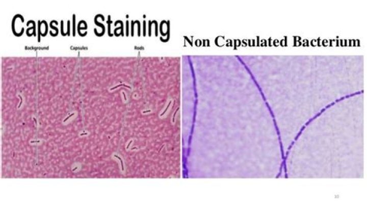
Bacterial capsules are composed of high-molecular-weight polysaccharides and/or polypeptides, and are associated with virulence and biofilm formation. Unfortunately, capsules do not stain well with crystal violet, methylene blue, or other simple stains.
What are two things that are stained in a capsule stain?
Capsule stain is a type of differential stain which involves the use of two stains; primary stain and the counterstain.
What are the two things that are stained in a capsule stain quizlet?
- Polypeptides.
- Polysaccharide.
- Both Polypeptides and Polysaccharide.
What is stained in a capsule stain?
Capsule stain is a type of differential stain which uses acidic and basic dyes to stain background & bacterial cells respectively so that presence of capsule is easily visualized. Capsule is synthesized in the cytoplasm and secreted to the outside of the cell where it surrounds the bacterium.Is capsule resistant to staining?
Capsular material is very moist (slimy) and any heating will cause it to shrink – it is for this reason that we will not heat fix the slide before staining. … As a general phenomenon, organisms with capsules tend to be more virulent presumably because of their resistance to phagocytosis and killing.
What type of staining is used for capsules and why?
Bacterial capsules are non-ionic, so neither acidic nor basic stains will adhere to their surfaces. Therefore, the best way to visualize them is to stain the background using an acidic stain and to stain the cell itself using a basic stain. We use India ink and Gram crystal violet.
What is a bacterial capsule composed of?
The capsule is composed of polysaccharides that cover the cell wall, which is made up of peptidoglycan and teichoic acid, characterizing the classic gram positive structure; It acts as the principal antiphagocytic and protective element that prevents access of the leukocytes to the underlying cell wall elements.
What color is the capsule following the capsule stain?
Selectively stains bacterial capsules. Capsule appears as a faint blue halo around a purple cell.Why is copper sulfate used in capsule staining?
In Anthony’s capsule stain, crystal violet is used as the primary stain, interacting with the protein material in the culture broth or added during the staining, and copper sulfate serves as the mordant. There is no additional negative stain.
Is Endospore stain a differential stain?The endospore stain is a differential stain used to visualize bacterial endospores. Endospores are formed by a few genera of bacteria, such as Bacillus . By forming spores, bacteria can survive in hostile conditions. Spores are resistant to heat, dessication, chemicals, and radiation.
Article first time published onWhy is the capsule stain not heat fixed quizlet?
Why Do You NOT Heat Fix A Capsule Stain Slide? Heat Fixing Causes The Cells To Shrink Leaving An Artificial White Halo Around Them That Could Be Mistaken For A Capsule. Gram Staining is A Differential Staining Method Used To Determine Between Gram Positive Or Gram Negative.
Which stain is used to stain the background in the capsule stain quizlet?
Use a basic dye (crystal violet, carbon fuchsin) to color the bacterial cell. The positive charge of the dye is attracted to the slightly negative charge of the cell, so it stains it. Uses an acidic dye (Congo Red) to stain the background of the slide while the specimens appears light or transparent.
How do you do a capsule stain?
- Prepare thin smears of bacterial culture on a microscope slide.
- Allow the smear to only air-dry. …
- Apply 1% crystal violet and allow it to remain on the slide for 2 minutes.
- With the slide over the proper waste container provided, gently wash off the crystal violet with 20% copper sulfate.
What color do endospores and bacteria stain?
Whereas the counterstain (safranin) is pink/reddish in color, the primary stain (malachite green) is green in color. Therefore, endospores will appear green in color while the vegetative cells will pink/reddish in color under the microscope.
What are most capsules made of?
Capsules are made up of gelatin (hard or soft) and nongelatin shells generally derived from hydrolysis of collagen (acid, alkaline, enzymatic, or thermal hydrolysis) from animal origin or cellulose based.
Why would a microorganism want to produce a capsule?
The capsule is considered a virulence factor because it enhances the ability of bacteria to cause disease (e.g. prevents phagocytosis). The capsule can protect cells from engulfment by eukaryotic cells, such as macrophages. A capsule-specific antibody may be required for phagocytosis to occur.
What is capsule microbiology?
Capsules are the outmost structures of bacterial and fungal cells. The capsules protect microbial cells from immune recognition and killing during infection of mammalian hosts. Except for the poly-γ-glutamate (PGA) capsule of Bacillus anthracis, other known capsules are all composed of polysaccharides.
What is capsule in prokaryotic cell?
Many prokaryotes have a sticky outermost layer called the capsule, which is usually made of polysaccharides (sugar polymers). The capsule helps prokaryotes cling to each other and to various surfaces in their environment, and also helps prevent the cell from drying out.
How is a bacterial capsule formed?
Bacterial capsules are formed primarily from long-chain polysaccharides with repeat-unit structures. A given bacterial species can produce a range of capsular polysaccharides (CPSs) with different structures and these help distinguish isolates by serotyping, as is the case with Escherichia coli K antigens.
How is the capsule stain used in clinical microbiology?
How is the capsule stain used in the clinical microbiology? … A capsule to a bacterium helps bacteria adhere to surfaces and resist flushing. Several bacteria that have capsules are klebsiella, pneumoniae, bacillus subtillus, and streptococcus pneumoniae.
What color will capsules appear with a Gram stain?
In a capsule stained microscope image, the bacterial cells will typically be stained purple, and the background of the slide should be darkly stained. Against this dark background, the capsules of the bacteria, if present, will appear as a clear halo around the cells.
Why is heat not used in capsule staining?
Do not heat-fix the smear because this can destroy the capsule and will also cause shrinkage of the bacteria, making a clear area around the cell that can mistakenly be identified as a capsule.
What does a bacterial capsule do?
Capsules can protect a bacterial cell from ingestion and destruction by white blood cells (phagocytosis). While the exact mechanism for escaping phagocytosis is unclear, it may occur because capsules make bacterial surface components more slippery, helping the bacterium to escape engulfment by phagocytic cells.
Does Klebsiella pneumoniae have a capsule?
The capsular polysaccharide and type 1 fimbriae are two of the major surface-located virulence properties associated with the pathogenesis of Klebsiella pneumoniae. The capsule is an elaborate polysaccharide matrix that encases the entire cell surface and provides resistance against many host defense mechanisms.
Why are capsule stains not rinsed with water?
How it works: Capsules are highly ordered polymers of sugars and proteins that surround some bacterial cells, and can be easily dislodged by heat or water. Accordingly, capsule stains are not heat-fixed, and water is never used to rinse.
What color do endospores and bacteria stain in an Endospore stain quizlet?
What colors do the endospores stain? The spores stain green, and the vegetative cells stain pink.
How endospore is stained?
The Schaeffer-Fulton method is the most commonly used endospore staining technique, which uses Malachite green as the primary stain. Once the endospore has absorbed the stain, it is resistant to decolorization, but the vegetative cell is easily decolorized with water (leaving the vegetative cells colorless).
What color is a negative Endospore stain?
Principle of Dorner’s method for staining endospores Since the counterstain nigrosin is negatively charged, bacterial cells don’t easily take up the counterstain. Therefore, vegetative cells appear colorless, endospores stain red, and the background is black.
What is the purpose of the capsule stain quizlet?
What is the purpose of a Capsule stain? differential stain used to detect cells capable of producting an extracelluar capsule (usually pathogenic).
What stain is added to the dried primary stain in a capsule stain quizlet?
The heat might alter the morphology of the bacteria. A capsule stain was performed, using crystal violet as a primary stain, followed by a water rinse, and then nigrosin as a counterstain. As you observe the slide through the microscope, you suspect you made an error.
Why are endospores resistant to heat and chemicals quizlet?
Why are endospores resistant to heat and chemicals? due to a tough outer coating made of a protein called keratin, its DNA protective proteins and its dehydrated state. … An endospore is a spore in a capsule that is resistant to harsh conditions which is alive but does not reproduce or produce ATP.
