Synovial fluid is produced by the synovium and is composed of water, inorganic salts and macromolecules, hyaluronic acid, lubricin and aggrecans, which contribute to the boundary lubrication.
What is synovial fluid made up of?
In normal/healthy joint, the synovial fluid, which consists of an ultrafiltrate of blood plasma and glycoproteins contains HA macromolecules of molar mass ranging between 6–10 mega Daltons (Praest et al., 1997). SF serves also as a lubricating and shock absorbing boundary layer between moving parts of synovial joints.
What does synovial fluid provide?
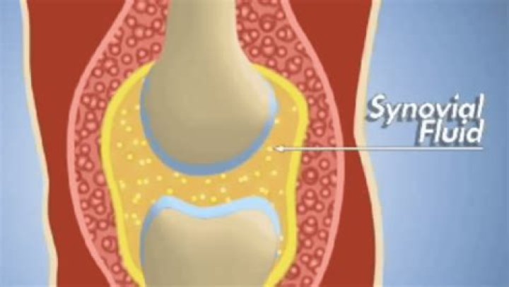
Synovial fluid, also known as joint fluid, is a thick liquid located between your joints. The fluid cushions the ends of bones and reduces friction when you move your joints.
What cells are found in synovial fluid?
Nucleated cells recognized frequently in synovial fluid include neutrophils, lymphocytes, monocytes, and macrophages [9]. These cells are seen in fluids from normal as well as diseased joints.What is the fluid inside the synovial membrane called?
secretion by synovial tissue secrete a viscous liquid called synovial fluid; this liquid contains protein and hyaluronic acid and serves as a lubricant and nutrient for the joint cartilage surfaces.
What are synovial macrophages?
Synovial macrophages are one of the resident cell types in synovial tissue and while they remain relatively quiescent in the healthy joint, they become activated in the inflamed joint and, along with infiltrating monocytes/macrophages, regulate secretion of pro-inflammatory cytokines and enzymes involved in driving the …
What are the components of a synovial joint?
Synovial joints share important structural components: subchondral bone, hyaline cartilage, a joint cavity, synovial lining, articular capsule, and supporting ligaments.
Does synovial fluid contain phagocytic cells?
Synovial fluid contains phagocytic cells that protect the cavity from invasion by microbes or other debris. A person who has been diagnosed with rheumatoid arthritis would be suffering loss of the synovial fluids. A ball-and-socket joint is a multiaxial joint.What is synovial fluid tested for?
A synovial joint fluid analysis is a group of tests your doctor can use to diagnose problems with your joints. Joint conditions like arthritis, gout, infections, and bleeding disorders can change how your synovial fluid looks and feels.
What releases synovial fluid?A joint’s synovial membrane produces substances called albumin and hyaluronic acid that give the synovial fluid its viscosity and slickness. In addition, synovial fluid delivers nutrients to the cartilage and removes waste from the cartilage. When a joint is at rest, cartilage absorbs some of the synovial fluid.
Article first time published onWhat's the fluid in your knee called?
Knee effusion, sometimes called water on the knee, occurs when excess fluid accumulates in or around the knee joint. Common causes include arthritis and injury to the ligaments or meniscus, which is cartilage in the knee.
How synovial fluid is formed?
Synovial fluid is formed through a serum ultrafiltration process by cells that form the synovial membrane (synoviocytes). Synovial cells also manufacture hyaluronic acid (HA, also known as hyaluronate), a glycosaminoglycan that is the major noncellular component of synovial fluid.
What is found in synovial fluid quizlet?
Synovial fluid is the same composition as plasma with what two substances added? A mucopolysaccharide containing hyaluronic acid and a small amount of protein. What is the name of the procedure used to collect joint fluid? Arthrocentesis.
What is synovial villi?
The synovial villi are small vascular processes developing from the synovial membrane.
What structure in the synovial joint produces synovial fluid?
The inner membrane of synovial joints is called the synovial membrane and secretes synovial fluid into the joint cavity. Synovial fluid is an ultrafiltrate from plasma, and contains proteins derived from the blood plasma and proteins that are produced by cells within the joint tissues.
What are the 7 structures of a synovial joint?
- Joint capsule. Sleeve-like extension of the periosteum of each of the articulating bones.
- Synovial Membrane. Moist, slippery membrane that lines the inner surface of the joint capsule.
- Articular Cartilage. …
- Joint Cavity. …
- Menisci (articulatin disks) …
- Ligaments. …
- Bursae. …
- Bony prominences.
What type of cartilage is found in synovial joints?
At synovial joints, the articular surfaces of bones are covered with smooth articular cartilage. This gives the bones of a synovial joint the ability to move smoothly against each other, allowing for increased joint mobility.
Does synovial fluid contain macrophages?
In contrast, macrophages are relatively more present in the synovial fluid in OA as compared to RA [21]. Furthermore, macrophages are also found to be the predominant inflammatory cell type in the synovial tissue in OA [12,22,23].
What are synovial tissues?
Synovial tissue is a highly specialized tissue that keeps the articular joint well lubricated, and at the same time provides nutrients to the articular surface. A joint needs a small amount of synovial fluid to work. Although the tissue structure is simple, its function is highly sophisticated.
What happens if you have too much synovial fluid?
As rheumatoid arthritis progresses, the synovium, which produces synovial fluid, swells and thickens, producing an excess of synovial fluid. This, in turn, leads to further swelling and inflammation which causes pain and stiffness in the joint.
What happens if you lose synovial fluid?
Since cartilage is porous, synovial fluid leaks out its holes every day. Permanent loss of this fluid results in a gradual decrease in cartilage thickness and increase in friction, which can lead to the joint degeneration of osteoarthritis, the most common form of arthritis.
What does abnormal synovial fluid mean?
Abnormal synovial fluid may be cloudy and thicker or thinner than normal fluid. Cloudiness could mean there are crystals, excess white blood cells, or microorganisms in the fluid. If you have gout, the fluid will contain crystals. Less stringiness in the fluid could signal inflammation.
Are mesothelial cells found in synovial fluid?
Synovial lining cells are indicated by the arrow in the image on the right. They resemble miniature mesothelial cells or small macrophages. They can be found singly or in clumps and can have “foamy”-looking cytoplasm.
What does protein in synovial fluid mean?
Synovial fluid protein levels greater than 2.5 g/dl are abnormal, and those greater than 4.5 g/dl indicate significant inflammation. In inflammatory effusions, a gram stain and culture are mandatory to rule out a septic joint as well as infection superimposed on another condition, such as rheumatoid arthritis.
What are neutrophils in synovial fluid?
Neutrophils are believed to play an important role in both the initiation and progression of RA, and large numbers of activated neutrophils are found within both synovial fluid (SF) and synovial tissue from RA joints.
Where is synovial fluid present?
Synovia or synovial fluid is a viscous, non-Newtonian fluid present in the cavities of synovial joints and has the consistency as that of an egg-white. It is a thick liquid that lubricates our joints and enables their smooth movement. They are found in our joints in the knees, hips, shoulders, feet, hands.
What foods increase synovial fluid?
- Dark, leafy vegetables.
- Foods rich in omega-3 fatty acids like salmon, mackerel, and flaxseeds.
- Anti-inflammatory foods rich in compounds like curcumin (found in turmeric)
- Foods high in antioxidants like onions, garlic, green tea, and berries.
- Nuts and seeds.
Where are menisci found?
The menisci sit between the tibia (lower leg bone) and the femur (thigh bone) and protect the lower part of the leg from the shock created by our body weight. The medial meniscus sits on the inside of the knee and the lateral meniscus sits on the outside of the knee.
How do I get rid of synovial fluid in my knee?
- R.I.C.E.—which stands for rest, ice, compression, and elevation—to relieve minor pain directly after an injury.
- Compression by gently wrapping the knee with elastic bandages.
- Over-the-counter nonsteroidal anti-inflammatory pain medication (NSAIDs), such as ibuprofen or naproxen.
- Physical therapy exercises.
What does it mean when the back of your leg hurts behind the knee?
Some of the most common causes of pain behind the knee (posterior knee pain) include, Baker’s cyst, arthritis, infection, injury, tumor, or deep vein thrombosis. Since the knee is the largest and most complex joint in the body, it makes sense that it might hurt sometimes.
Is walking good for a swollen knee?
You may worry that a walk will put extra pressure on your joints and make the pain worse. But it has the opposite effect. Walking sends more blood and nutrients to your knee joints. This helps them feel better.
