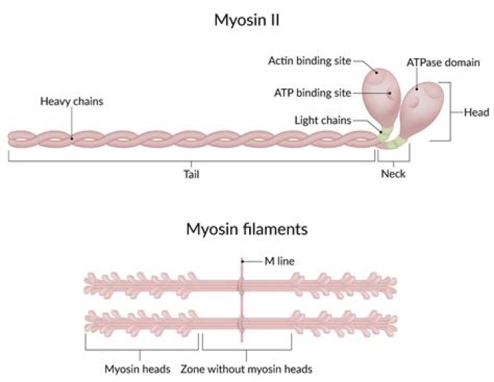
The type of myosin present in muscle (myosin II) is a very large protein (about 500 kd) consisting of two identical heavy chains (about 200 kd each) and two pairs of light chains (about 20 kd each) (Figure 11.22). Each heavy chain consists of a globular head region and a long α-helical tail.
What color is actin and myosin?
Sarcomeres contain two distinct types of bands. These are the dark-colored A bands, which contain thick, myosin filaments, and the I bands, which have a lighter color and contain only thin, actin filaments.
Is myosin thick or thin?
The myofibrils are made up of thick and thin myofilaments, which help give the muscle its striped appearance. The thick filaments are composed of myosin, and the thin filaments are predominantly actin, along with two other muscle proteins, tropomyosin and troponin.
What is myosin seen in?
Where Is Myosin Found? In both eukaryotic cells, cells that have membrane-bound organelles and a nucleus, and prokaryotic cells, cells that lack a nucleus and membrane-bound organelles, we can find myosin. It exists as a filament inside of the cell.Is myosin dark or light?
Dark bands contain actin and myosin filaments side by side. Light bands contain only actin filaments. Many students have limited opportunities to develop scientific expertise.
Where are myosin filaments located?
The actin filaments are attached at their plus ends to the Z disc, which includes the crosslinking protein α-actinin. The myosin filaments are anchored at the M line in the middle of the sarcomere.
Is myosin fibrous or globular?
Myosin is therefore unusual in that it is both a fibrous protein, and a globular enzyme.
What is the difference between myosin and actin?
The main difference between actin and myosin is that actin is a protein that produces thin contractile filaments within muscle cells, whereas myosin is a protein that produces the dense contractile filaments within muscle cells.Are actin and myosin Myofilaments?
Myofilaments are the two protein filaments of myofibrils in muscle cells. The two proteins are myosin and actin and are the contractile proteins involved in muscle contraction. The two filaments are a thick one composed mostly of myosin, and a thin one composed mostly of actin.
What are myosin filaments?Myosin filaments (also called thick filaments) are key components of muscle and non-muscle cells. In striated muscle, they overlap with thin (actin-containing) filaments in an orderly array, making a repeating pattern of sarcomeres, the basic units of contraction [1] (Figure 1a).
Article first time published onWhat organisms contain myosin?
Organisms on the branch including amoebas, yeast and animals have genes for myosins types I, II, and V, but myosin genes diversified in animals, so humans have 40 myosin genes from 13 classes. Gene duplications gave rise to multiple isoforms within most classes of myosin.
Is myosin found in cytoplasm?
Although originally characterized as a cytoplasmic protein, myosin of various classes also performs key functions in the nucleus.
What do Myofilaments look like?
Thick myofilaments are comprised of around 300 spirally arranged protein molecules called myosin. Each myosin molecule is composed of two heavy chains and four light chains.
Is myosin a substance?
myosin | chemical compound | Britannica.
How is myosin synthesized?
The steps in its synthesis are; Transcription, where the nucleotide sequence in a myosin gene is copied to form mRNA. mRNA undergoes modification to safely reach ribosomes in the cytoplasm. Translation, during which amino acids are arranged in a sequence according to mRNA to make myosin molecule.
What type of muscle never gets tired?
Cardiac muscles are involuntary muscles found only in the heart. Cardiac muscles do not get tired.
Are I bands light or dark?
The light bands are called I bands and contain only thin filaments. The dark bands are called A bands and contain thick and thin filaments, with the thick filaments running the entire length of the A band.
What is the function of myosin tail?
Myosin II is a motor protein with two heads and an extended tail that plays an essential role in cell motility. Its active form is a polymer (myosin filament) that pulls on actin to generate motion.
Is insulin fibrous or globular?
For example, insulin is a ball-shaped, globular protein that contains both hydrogen bonds and disulfide bonds that hold its two polypeptide chains together. Silk is a fibrous protein that results from hydrogen bonding between different β-pleated chains.
Do humans have myoglobin?
Myoglobin is found in your heart and skeletal muscles. There it captures oxygen that muscle cells use for energy. When you have a heart attack or severe muscle damage, myoglobin is released into your blood. Myoglobin increases in your blood 2 to 3 hours after the first symptoms of muscle damage.
Is albumin is an example of fibrous protein?
Such proteins are insoluble in water. Some examples of fibrous proteins include: Keratin, Myosin, collagen etc. … Such proteins are soluble in water and some examples include: Haemoglobin, Albumin, Insulin etc.
What would happen if myosin is damaged?
Because of muscle weakness, affected individuals may start walking later than usual and have a waddling gait, trouble climbing stairs, and difficulty lifting the arms above shoulder level. Muscle weakness also causes some affected individuals to have trouble breathing.
Is myosin part of the cytoskeleton?
Of the three types of protein fibers in the cytoskeleton, microfilaments are the narrowest. … For one, they serve as tracks for the movement of a motor protein called myosin, which can also form filaments. Because of its relationship to myosin, actin is involved in many cellular events requiring motion.
What is the function of actin filaments?
Actin filaments are particularly abundant beneath the plasma membrane, where they form a network that provides mechanical support, determines cell shape, and allows movement of the cell surface, thereby enabling cells to migrate, engulf particles, and divide.
How does myosin bind to actin?
Myosin binds to actin at a binding site on the globular actin protein. Myosin has another binding site for ATP at which enzymatic activity hydrolyzes ATP to ADP, releasing an inorganic phosphate molecule and energy. ATP binding causes myosin to release actin, allowing actin and myosin to detach from each other.
What are transverse tubules made of?
Structure. T-tubules are tubules formed from the same phospholipid bilayer as the surface membrane or sarcolemma of skeletal or cardiac muscle cells.
Where is Epimysium found?
Epimysium (plural epimysia) (Greek epi- for on, upon, or above + Greek mys for muscle) is the fibrous tissue envelope that surrounds skeletal muscle. It is a layer of dense irregular connective tissue which ensheaths the entire muscle and protects muscles from friction against other muscles and bones.
Whats bigger actin or myosin?
Actin proteins are found in both A and I bands of the sarcomere. Myosin proteins are found only in the A bands of the sarcomere. These are shorter (2-2.6 µm in length) and thinner (0.005 µm in diameter). These are longer (4-5 µm in length) and thicker (0.01 µm in diameter).
Is myosin a structural protein?
Structural proteins include the contractile proteins (actin and myosin), the major regulatory proteins (troponin and tropomyosin), the minor regulatory proteins (M-protein, C-protein, F-protein, I-protein, and actinins), and the scaffold proteins (connectin, desmin, and Z-protein).
When actin and myosin interact What happens?
The actin–myosin interaction produces two types of movements: force generation between actin filaments leading to contractions, such as in muscle contraction, cell motility, and cytokinesis; and transport of subcellular organelles and macromolecular complexes by myosin motors along actin filaments.
How many amino acids are in myosin?
In myosin II molecules, there is usually a 26 amino acid separation between the start of IQ motifs, but in unconventional myosins, the separation can be between 23 and 26 residues [32]. For example, in myosin V molecules, the six IQ motifs are separated by an alternating pattern of 23 and 25 residues.
