The extraembryonic mesoderm supports the epithelium of the amnion
What happens extraembryonic Coelom?
It communicates temporarily with the coelomic cavity within the embryo (peritoneal cavity). Late in pregnancy it becomes almost entirely obliterated by the growth of the amnion, which fuses with the chorion.
Between which layers is the extraembryonic mesoderm located?
Between which two layers is the extraembryonic mesoderm located? E. The extraembryonic mesoderm is derived from the epiblast and is located between the exocoelomic membrane and the cytotrophoblast.
Is extra-embryonic mesoderm primary mesoderm?
Extra-embryonic mesoderm Extraembryonic somatic mesoderm lines the cytotrophoblast and covers the amnion is. Extraembryonic somatic mesoderm also forms the connecting stalk that is the primordium of the umbilical cord. Extraembryonic visceral mesoderm covers the yolk sac.What happens to the Embryoblasts?
The embryoblast is the source of embryonic stem cells and gives rise to all later structures of the adult organism. The trophoblast combines with the maternal endometrium to form the placenta in eutherian mammals.
What are extraembryonic cells?
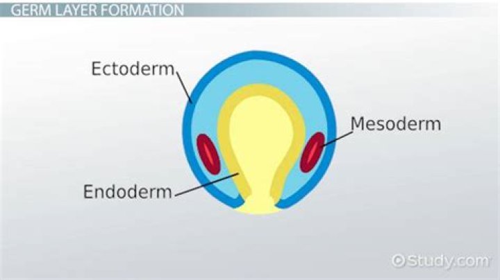
All amniotes contain the following four extraembryonic components: the amnion, chorion, yolk sac, and allantois (Fig. … Like the intraembryonic tissues, these extraembryonic tissues are composed of cells representing the three germ layers: ectoderm, mesoderm, and endoderm.
How does the blastocyst develop?
In humans, blastocyst formation begins about 5 days after fertilization when a fluid-filled cavity opens up in the morula, the early embryonic stage of a ball of 16 cells. … About seven days after fertilization, the blastocyst undergoes implantation, embedding into the endometrium of the uterine wall.
Where does the amnion come from?
The amnion arises from the epiblast cells of the blastocyst and grows to surround the developing embryo, creating a fluid-filled cavity. Thus, throughout prenatal life, humans are surrounded by AF. This fluid serves many functions critical for prenatal development.What is extraembryonic ectoderm?
The extraembryonic ectoderm (ExE) is formed following implantation as cells from the polar trophectoderm proliferate. … The ExE later forms the chorionic ectoderm and is the source of undifferentiated trophoblast stem cells, which, in the mouse embryo, contribute to the ectoplacental cone and to the mature placenta.
Is a yolk sac diverticulum It is derived from extraembryonic mesoderm?It is also known as the exocoelomic cavity. Secondary yolk sac: this structure is formed when the extraembryonic mesoderm separates to form the extraembryonic coelom; cells from the mesoderm pinch off an area of the yolk sac, and what remains is the secondary yolk sac.
Article first time published onWhat does Blastocoel become?
The blastocoel is a fluid filled cavity, or space, in the developmental stage known as the blastula, which in mammals is called a blastocyst. … These aid in the growth and change of the cells in the blastocoel that will become the embryo.
What does the connecting stalk become?
The connecting stalk, which is the precursor of the umbilical cord, is formed by mesenchymal cells, and it connects the amnion cavity and the extracoelomic cavity.
What type of mesoderm gives rise to muscles in the limb bone and cartilage?
Somites form (1) the cartilage of the vertebrae and ribs, (2) the muscles of the rib cage, limbs, and back, and (3) the dermis of the dorsal skin.
What does the Sclerotome become?
The sclerotome forms the vertebrae and the rib cartilage and part of the occipital bone; the myotome forms the musculature of the back, the ribs and the limbs; the syndetome forms the tendons and the dermatome forms the skin on the back.
What is morula stage?
An early stage in post-fertilization development when cells have rapidly mitotically divided to produce a solid mass of cells (16 or more) with a “mulberry” appearance is called the morula stage. The morula stage is the final stage prior to the formation of a fluid-filled cavity called the blastocoel cavity.
What does the ICM turn into?
Inner cell massFMA86557Anatomical terminology
Why is the blastocyst important?
Significance of Blastocyst The blastocyst is the highest degree of development that an embryo can reach in vitro. … In in vitro fertilization (IVF), the blastocyst culture is important to increase the success rate of IVF because of better embryo selection after better genomic activation and endometrial receptivity.
What are the 4 stages of embryonic development?
- 1.1 Fertilization.
- 1.2 Cleavage.
- 1.3 Blastulation.
- 1.4 Implantation.
- 1.5 Embryonic disc.
What is morula and blastocyst?
A morula is distinct from a blastocyst in that a morula (3–4 days after fertilization) is a mass of 16 totipotent cells in a spherical shape whereas a blastocyst (4–5 days after fertilization) has a cavity inside the zona pellucida along with an inner cell mass.
What do extraembryonic membranes become?
In placental mammals, the extraembryonic membranes form a placenta and umbilical cord, which connect the embryo to the mother’s uterus in a more elaborate and efficient way. The blood supply of the developing fetus is continuous with that of the placenta.
What are the extraembryonic membranes and their functions?
The thin layers of tissue that surround the developing embryo. There are four extra-embryonic membranes commonly found in VERTEBRATES, such as REPTILES; BIRDS; and MAMMALS. They are the YOLK SAC, the ALLANTOIS, the AMNION, and the CHORION. These membranes provide protection and means to transport nutrients and wastes.
How extraembryonic membranes are formed?
Another extraembryonic membrane formed from the inner cell mass, the amnion, then grows over the forming embryo (Figure 10.4). The amniotic cavity becomes filled with amniotic fluid. The amnion is an important extraembryonic membrane throughout development.
Where does extraembryonic ectoderm come from?
The tissues colored green (the extraembryonic ectoderm and ectoplacental cone (ec)) are derived from the trophectoderm. The tissues in yellow are derived from the primitive endoderm and epiblast cells that passed through the streak, generating the definitive endoderm.
What is extra embryonic endoderm?
The extraembryonic endoderm is a derivative of the hypoblast cells that migrate into the blastocyst cavity (beginning on day 8 of human embryonic development), and line the cavity, giving rise to the primary and definitive yolk sacs. The extraembryonic endoderm fills the remaining cavity of the blastocyst.
What does Allantois become?
The human allantois is a caudal out-pouching of the yolk sac, which becomes surrounded by the mesodermal connecting stalk known as the body-stalk. … The embryonic allantois becomes the fetal urachus, which connects the fetal bladder (developed from cloaca) to the yolk sac.
What is the main function of amnion?
The amnion is a thin, tough membrane that protects a developing child. It allows for nutrients to reach the fetus and wastes to be removed. Amniotic fluid is found inside of the amnion and will provide protection for the developing child until it is time for the pregnancy to end.
Where is the mesoderm?
The mesoderm is the middle layer of the three germ layers that develop during gastrulation in the very early development of the embryo of most animals. The outer layer is the ectoderm, and the inner layer is the endoderm. The mesoderm forms mesenchyme, mesothelium, non-epithelial blood cells and coelomocytes.
What is the role of amnion in chordate evolution?
The amnion acts to protect the developing embryo against mechanical shock. The chorion and the allantois are involved in gas exchange of oxygen and carbon dioxide, while the allantois transports nutrients to, and waste from, the embryo.
Why does the yolk sac not develop?
A blighted ovum, also called an anembryonic pregnancy, occurs when an early embryo never develops or stops developing, is resorbed and leaves an empty gestational sac. The reason this occurs is often unknown, but it may be due to chromosomal abnormalities in the fertilized egg.
What is the yolk sac derived from?
The yolk sac is formed from hypoblast endoderm and extraembryonic mesoderm. The hypoblast separates from the inner surface of the embryonic disc in early blastocyst stage, forming an endodermal tube within the trophoblast tube. The hypoblast tube is invested with splanchnic mesoderm after its formation and splitting.
Which of the following are the extra embryonic membranes formed during chick development?
During the chick embryonic development (ED), three extraembryonic membranes are formed; (1) the yolk sac membrane, (2) the amnion, and (3) the chorioallantoic membrane (CAM).
