The conus medullaris give rise to the lumbar sympathetic, sacral somatic and sacral parasympathetic nerves which continue downward within the cauda equina
What happens if the conus medullaris is damaged?
Following a spinal cord injury, symptoms of conus medullaris syndrome develop rapidly and on both sides of the body by presenting: Weakness or tingling in your lower limbs. Sexual dysfunction. Incontinence.
What symptoms results in conus medullaris injury?
- Severe back pain.
- Strange or jarring sensations in the back, such as buzzing, tingling, or numbness.
- Bowel and bladder dysfunction, such as difficulty controlling your elimination functions.
- Sexual dysfunction.
- Weakness, numbness, or tingling in your lower limbs.
What is the significance of the conus medullaris and cauda equina?
Function The conus medullaris and cauda equina give rise to spinal nerves L2-S5 and the coccygeal nerve. The anterior rami of these spinal nerves contribute to the lumbar and sacral plexuses, which provide motor and sensory innervation to the entire lower limb, pelvic and perineal regions.What extends from the conus medullaris?
At the distal end of the cord, many spinal nerves extend beyond the conus medullaris to form a collection that resembles a horse’s tail. This is the cauda equina.
What is a conus?
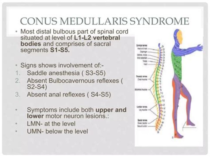
The conus medullaris (Latin for “medullary cone”) or conus terminalis is the tapered, lower end of the spinal cord. It occurs near lumbar vertebral levels 1 (L1) and 2 (L2), occasionally lower. … The filum terminale provides a connection between the conus medullaris and the coccyx which stabilizes the entire spinal cord.
Where should conus medullaris terminate?
Conclusions: The CM terminates most commonly at the L1-2 disc space and in the absence of tethering, the CM virtually never ends below the mid-body of L2. A CM that appears more caudal on neuroimages should be considered tethered.
Is conus medullaris upper motor neuron?
The symptoms and signs of cauda equina syndrome tend to be mostly lower motor neuron (LMN) in nature, while those of conus medullaris syndrome are a combination of LMN and upper motor neuron (UMN) effects (see Table 1, below).What is the conus medullaris made of?
The terminal portion of the spinal cord in the lumbar region is cone-shaped and is called the conus medullaris. The conus medullaris is made up of several neurons (nerve cells) and has 3 protective layers. Starting from the outermost layer, these are the dura mater, arachnoid mater, and pia mater.
What is conus medullaris at T12?The conus medullaris, the termination of the spinal cord, is located anywhere between T12 and lower L2. 9. The conus is the last segment of the cord from which nerve roots arise; none arise from the filum.
Article first time published onWhat is a low lying Conus?
Low-lying conus medullaris: It refers to a low position of a normal-appearing conus medullaris with respect to the vertebral level. It is usually located between the T12–L1 and L1–L2 disk level; however, in 6.4% of population it can be found between the upper and middle third of L2.
Where does the conus medullaris start?
The lowermost tapering extremity of the spinal cord is called the conus medullaris, which is around the first or second lumbar vertebra and can sometimes be lower.
Where does conus medullaris end in adults?
The conus medullaris most commonly terminates at the L1/2 intervertebral disc level in children and adults 1-3. Extending from the conus is a delicate strand of fibrous tissue called the filum terminale that acts to give longitudinal support to the cord.
What level does the conus medullaris lie?
Conus medullaris ended at the level between the first and second lumbar vertebrae (L1–L2) in 88% and 78% of preterm and term babies, respectively. In the preterm group three patients (7.2%) had conus medullaris between the second and third lumbar vertebrae (L2–L3) and in one patient (2.4%) it was at the L4–L5 level.
What is the Conus equina?
The collection of nerves at the end of the spinal cord is known as the cauda equina, due to its resemblance to a horse’s tail. The spinal cord ends at the upper portion of the lumbar (lower back) spine.
What is Conus and Epiconus?
The conus medullaris contains neural tissue from sacral cord segments S2, S3, S4, S5 and coccygeal 1. Superiorly, the epiconus extends from lumbar cord segments L3, L4, L5 and sacral cord segment S1. This usually corresponds to vertebral levels T12 and L1.
Where does conus medullaris end in newborns?
The caudal end of the spinal cord corresponds to the conus medullaris (,Fig 4), which continues into the filum terminale. In healthy newborns, the tip of the conus medullaris is located between L1 and L2. The tip should not be positioned below L2-3 (,8).
What is Conus shipping?
CONUS is shorthand for the continental United States (i.e., the 49 states, including Alaska and the District of Columbia and excluding Hawaii, on the North American continent). … OCONUS adds a “O” for “Outside” and refers to transportation logistics and shipping services outside the continental United States.
What does Hydrosyringomyelia mean?
Syringomyelia is a long-term condition that causes fluid-filled cysts, which doctors call “syrinx,” to form inside your spinal cord. You may also hear it called hydromyelia, syringohydromyelia, or Morvan disease. You may not have symptoms, or even be aware that you have it.
What do dorsal roots do?
Dorsal nerve roots carry sensory neural signals to the central nervous system (CNS) from the peripheral nervous system (PNS). … Until recently, the dorsal root ganglion has been considered a passive organ that metabolically assists functions and pathways between the PNS and CNS.
Why is cauda equina an emergency?
When the Cauda Equina nerves are compressed this normally results in what are commonly referred to as ‘red flag’ symptoms. Cauda Equina Syndrome is a medical emergency because delayed decompression surgery can result in lifelong disability.
Can you live with cauda equina syndrome?
Although cauda equina syndrome is not a fatal condition, it can cause severe neurological damage. If the condition is not treated quickly enough, this damage may be irreversible, meaning a patient will not make a full recovery.
What are the first signs of cauda equina?
- Lower limb weakness and intermittent changes in sensation, such as numbness.
- “Saddle anesthesia” – loss or diminished sensation in areas where a person would sit on a saddle.
- Urinary and/or bowel problems, such as retention or incontinence.
Is the cauda equina inferior to the conus medullaris?
Cauda equina and filum terminale seen from behind. … The cauda equina occupies the lumbar cistern, a subarachnoid space inferior to the conus medullaris.
Is cauda equina an emergency?
Cauda equina syndrome is a rare disorder that usually is a surgical emergency. In patients with cauda equina syndrome, something compresses on the spinal nerve roots. You may need fast treatment to prevent lasting damage leading to incontinence and possibly permanent paralysis of the legs.
What is tether cord?
What is a tethered spinal cord? A tethered spinal cord is a spinal cord that is pulled down and stuck, or fixed, to the spinal canal. The spinal cord normally floats free inside the spinal canal. As a child grows, the spinal cord must be able to move freely inside the spinal canal.
What structure is located inferior to the conus medullaris of the spinal cord?
At the bottom of the spinal cord (conus medullaris) is the cauda equina, a collection of nerves that derives its name from the Latin translation of “horse’s tail” (early anatomists thought the collection of nerves resembled a horse’s tail).
What is Brown Séquard syndrome?
Brown-Séquard syndrome is a rare spinal disorder that results from an injury to one side of the spinal cord in which the spinal cord is damaged but is not severed completely. It is usually caused by an injury to the spine in the region of the neck or back.
What is the etiology of spinal Dysraphism?
The causes of spinal dysraphism are not yet completely understood. Genetic and environmental factors both seem to play a role. The spinal cord arises very early in fetal development–in the first several weeks of gestation. Many forms of spinal dysraphism develop during this time.
What is a Filar cyst?
A filar cyst is an incidental finding on neonatal lumbar sonography located in the filum terminale of the spinal cord. It is considered a normal variant and is often confused for a ventriculus terminalis, a smooth dilated cavity of the central canal, located within the conus medullaris.
What does low lying spinal cord mean?
A low-lying spinal cord (LLC) is defined as the conus medullaris ending below the L2 vertebrae. (1,2) An LLC is usually abnormally fixed to a caudal structure such as a lipoma or scar, which limits caudal-cranial movement. (3) This may be attributed to tethering of the spinal cord.
