The capsule is seen as a clear halo around the microorganism against the black background. This method is used for demonstrating Cryptococcus. The background will be dark (color of India ink). The bacterial cells will be stained purple (bacterial cells take crystal violet-basic dyes as they are negatively charged).
What color is capsule after staining?
Capsules appear colourless with stained cells against dark background. Capsules are fragile and can be diminished, desiccated, distorted, or destroyed by heating.
What would a capsule look like in a Gram stain?
In a capsule stained microscope image, the bacterial cells will typically be stained purple, and the background of the slide should be darkly stained. Against this dark background, the capsules of the bacteria, if present, will appear as a clear halo around the cells.
What color is a capsule at the end of the capsule stain What color is the bacteria quizlet?
Capsule appears as a faint blue halo around a purple cell. Can be easily dislodged by heat or water.What are two things that are stained in a capsule stain?
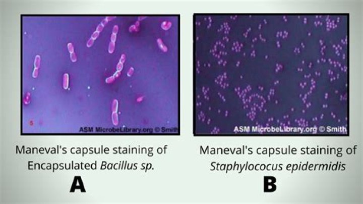
Capsule stain is a type of differential stain which involves the use of two stains; primary stain and the counterstain.
What is a capsule quizlet?
Capsules are solid dosage forms in which one or more medicinal or inert substances (as powder, compact, beads, or granulation) are enclosed within a small gelatin shell.
What color are endospores following the spore stain?
Whereas the counterstain (safranin) is pink/reddish in color, the primary stain (malachite green) is green in color. Therefore, endospores will appear green in color while the vegetative cells will pink/reddish in color under the microscope.
What are the two things that are stained in a capsule stain quizlet?
- Polypeptides.
- Polysaccharide.
- Both Polypeptides and Polysaccharide.
What color is a capsule at the end of the capsule stain What color is the bacteria?
A. The capsule is seen as a clear halo around the microorganism against the black background. This method is used for demonstrating Cryptococcus. The background will be dark (color of India ink). The bacterial cells will be stained purple (bacterial cells take crystal violet-basic dyes as they are negatively charged).
Is Gram negative pink or purple?Gram negative organisms are Red. Hint; Keep your P’s together; Purple is Positive. Gram stains are never pink they are red or purple so you don’t destroy the rule; keep your P’s together. In microbiology bacteria have been grouped based on their shape and Gram stain reaction.
Article first time published onWhat type of staining is Gram staining?
The Gram stain, the most widely used staining procedure in bacteriology, is a complex and differential staining procedure. Through a series of staining and decolorization steps, organisms in the Domain Bacteria are differentiated according to cell wall composition.
What is a bacterial capsule made of?
The capsule is composed of polysaccharides that cover the cell wall, which is made up of peptidoglycan and teichoic acid, characterizing the classic gram positive structure; It acts as the principal antiphagocytic and protective element that prevents access of the leukocytes to the underlying cell wall elements.
What type of staining is used for capsules and why?
Bacterial capsules are non-ionic, so neither acidic nor basic stains will adhere to their surfaces. Therefore, the best way to visualize them is to stain the background using an acidic stain and to stain the cell itself using a basic stain. We use India ink and Gram crystal violet.
How are capsules formed?
Bacterial capsules are formed primarily from long-chain polysaccharides with repeat-unit structures. A given bacterial species can produce a range of capsular polysaccharides (CPSs) with different structures and these help distinguish isolates by serotyping, as is the case with Escherichia coli K antigens.
How is the capsule stain used in clinical microbiology?
How is the capsule stain used in the clinical microbiology? … A capsule to a bacterium helps bacteria adhere to surfaces and resist flushing. Several bacteria that have capsules are klebsiella, pneumoniae, bacillus subtillus, and streptococcus pneumoniae.
What color do endospores and bacteria stain in an endospore stain quizlet?
What colors do the endospores stain? The spores stain green, and the vegetative cells stain pink.
Why do endospores stain green?
Because of their tough protein coats made of keratin, spores are highly resistant to normal staining procedures. The primary stain in the endospore stain procedure, malachite green, is driven into the cells with heat.
What does a positive endospore stain look like?
The presence of endospores in a bacterial culture can be detected by staining with malachite green. Because the endospore coat is so tough, steam is used to enable dye penetration. After washing, only the endospores will retain the primary stain Malachite green.
What is a capsule composed of quizlet?
What are capsules composed of? Majority of bacterial capsules are composed of polysaccharides. Some are composed of proteins or protein-carbohydrate combinations.
What is the significance of Congo Red in capsule staining How does it help visualize capsules under a microscope?
The positive charge of the dye is attracted to the slightly negative charge of the cell, so it stains it. Uses an acidic dye (Congo Red) to stain the background of the slide while the specimens appears light or transparent.
Which of the following capsules is the largest?
#000 capsules sometimes referred to as “triple zero” are the largest size of standard capsules.
What is the purpose of the capsule stain quizlet?
What is the purpose of a Capsule stain? differential stain used to detect cells capable of producting an extracelluar capsule (usually pathogenic).
What color is Nigrosin?
Nigrosin, an aqueous blue-black acid dye of the azine series, in conjunction with Biebrich scarlet, orange G and formic acid as mordant, it is found an excellent triple panchromatic rapid stain for histological purposes.
What is the role of the Congo Red in a capsule stain?
Congo red does not penetrate the capsule and provides a colored background. The sample is e then allowed to air dry. … The acetic acid lowers the pH in the sample and causes the Congo red to change from red to blue. The acid fuchsin penetrates through the capsule and stains the cell a bright red.
What stain is added to the dried primary stain in a capsule stain?
In Anthony’s capsule stain, crystal violet is used as the primary stain, interacting with the protein material in the culture broth or added during the staining, and copper sulfate serves as the mordant. There is no additional negative stain.
Which Dye did you first use in the capsule stain procedure?
a capsule stain was performed, using crystal violet as a primary stain, followed by a water rinse, and then nigrosine as a counterstain.
What kind of stain is most commonly used to visualize the capsule of Cryptococcus?
To visualize the capsule perimeter, we used India ink staining, which is excluded by the capsule, and the capsule can be exhibited by a halo around the cells.
What color are gram positive cells stained in the capsule stain what about Gram negative cells?
At the end of the gram staining procedure, gram positive cells will be stained a purplish-blue color. Gram negative cells also take up crystal violet, and the iodine forms a crystal violet-iodine complex in the cells as it did in the gram positive cells.
What color would human cells stain in the acid fast stain and why?
Because of its unique cell wall, when it is stained by the acid-fast procedure, it will resist decolorization with acid-alcohol and stain red, the color of the initial stain, carbol fuchsin.
What is the primary or negative stain used in the capsule stain quizlet?
The heat might alter the morphology of the bacteria. A capsule stain was performed, using crystal violet as a primary stain, followed by a water rinse, and then nigrosin as a counterstain.
Is Gram negative pink?
If the bacteria stays purple, they are Gram-positive. If the bacteria turns pink or red, they are Gram-negative.
