LocationMarkingLarge projection located lateral to headGreater tubercleLateral to the humeral head on anterior surfaceLesser tubercleGroove located between the greater and lesser tuberclesIntertubercular sulcus (groove)Roughened area located on the lateral aspect of the shaft of the humerusDeltoid tuberosity
What are bone markings?
Bone markings are projections and depressions found on bones, which help us to identify the location of other body structures, such as muscles. Their importance comes when we try to describe the shape of the bone or to understand how the muscles, ligaments and other structures affect this bone and vice versa.
Which of the following is a bone marking of the humerus quizlet?
Which of the following is a bone marking of the humerus? The deltoid tuberosity is a marking of the humerus. It is a roughened site where attachment to the deltoid muscle of the shoulder occurs.
What bone markings are part of the shoulder?
The bones of the shoulder consist of the humerus (the upper arm bone), the scapula (the shoulder blade), and the clavicle (the collar bone).What are the 9 bone markings?
- foramen. an opening through which blood vessels or nerves pass.
- meatus. a tubelike passageway running within a bone.
- paranasal sinus. an air-filled, mucosa lined, cavity within a bone connected to the nasal cavity.
- groove or sulcus. …
- fossa. …
- condyle. …
- head. …
- tubercle.
What are the two categories of bone markings Nasm?
- long.
- short.
- flat.
- irregular.
- sesamoid.
What is a tubercle bone marking?
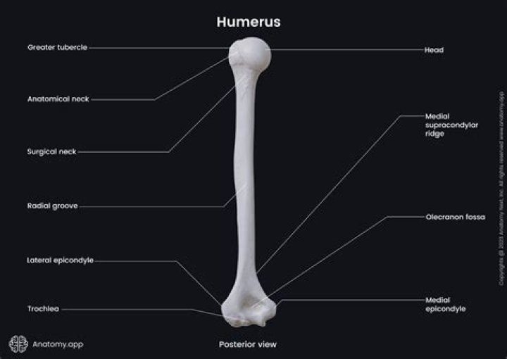
Tubercle – A small, rounded prominence where connective tissues attach. Examples include the greater and lesser tubercle of the humerus.
What type of bone is the humerus?
Your humerus is classified as a long bone. Other types of long bones include the radius and ulna in your forearm and the femur in your upper leg. Speaking of long, the humerus is the longest bone in your arm.What causes bone markings?
As with the other markings, their size and shape reflect the size of the vessels and nerves that penetrate the bone at these points. Figure 7.2. 1 – Bone Features: The surface features of bones depend on their function, location, attachment of ligaments and tendons, or the penetration of blood vessels and nerves.
Is Trochlea a bone marking?Trochlea (Trochlea humeri) is a pulley-shaped formation located medial to the capitulum. This region articulates with the trochlear notch of the ulna bone at the elbow joint.
Article first time published onWhat portion of the sternum is identified by the letter A?
Terms in this set (42) What portion of the sternum is identified by the letter A? The manubrium is the superior portion of the sternum.
Which of the following is a bone marking described as a round or oval?
Foramen: Round or oval opening through a bone. 3.
What is the proximal end of humerus?
At its proximal end is the head of the humerus. This is the large, round, smooth region that faces medially. The head articulates with the glenoid cavity of the scapula to form the glenohumeral (shoulder) joint. The margin of the smooth area of the head is the anatomical neck of the humerus.
What bone marking is an outgrowth from a bone?
Common Terms Used For Features of Bones (and other aspects of anatomy)TermDescriptionExampleProtuberanceA bony outgrowth or protruding partMental protuberance of the chinSpineA sharp, slender, or narrow processSpine of the scapulaTrochanterTwo massive processes unique to the femur
Where is the periosteum located?
The periosteum is a thin membrane on the outside of your bones. It serves to protect your bones but also has the ability to help them heal. It can even help your body grow new bone when damage occurs.
What is the Forum Magnum?
The foramen magnum (Latin: great hole) is a large, oval-shaped opening in the occipital bone of the skull. It is one of the several oval or circular openings (foramina) in the base of the skull. … It also transmits the accessory nerve into the skull. The foramen magnum is a very important feature in bipedal mammals.
What is the difference between tubercle and tuberosity?
The main difference between tuberosity and tubercle is that tuberosity refers to a slightly larger lump on bones, but tubercle refers to the smaller lump. Some of the examples of tuberosity are the greater tuberosity of the humerus and the ischial tuberosity of the hip bone.
What markings found on bones are indicative of nerve pathways?
Terms in this set (144) What markings found on bones are indicative of nerve pathways? Ridges. Foramina.
What bone marking is a small rounded bony projection?
ABTuberclesmall rounded projection or processEpicondyleraised area on or above a condyleProcessprominence or projectionHeadbony expansion carried on a narrow neck
What are mechanoreceptors Nasm?
Mechanoreceptors: any of the sense organs that respond to vibration, stretching, pressure, or other mechanical stimuli. Muscle Spindles: a proprioceptor that conveys information on the state of muscle stretch or length, important in the reflex mechanism that maintains body posture.
At what age is total peak bone mass reached Nasm?
By age 18, skeletal growth is nearly complete, with minor accumulations in bone density occurring until about age 30. At that point, bones have reached their maximum strength and density, known as peak bone mass. In women, there tends to be minimal change in total bone mass between age 30 and menopause.
Are tendons?
A tendon is a fibrous connective tissue that attaches muscle to bone. Tendons may also attach muscles to structures such as the eyeball. A tendon serves to move the bone or structure.
What bone makes up the upper arm?
Your arm is made up of three bones: the upper arm bone (humerus) and two forearm bones (the ulna and the radius).
Why do bones have openings?
A projection is an area of a bone that projects above the surface of the bone. … A hole is an opening or groove in the bone that allows blood vessels and nerves to enter the bone. As with the other markings, their size and shape reflect the size of the vessels and nerves that penetrate the bone at these points.
What are bone features?
Information. Most bones have some combination of bumps, ridges, projections, depressions, cavities, and holes in them that help them carry out their functions. These are where other structures like muscles, blood vessels and nerves, or other bones are attached to or articulate with or travel through the bone.
What is the top of the humerus called?
The head (caput humeri), is nearly hemispherical in form. It is directed upward, medialward, and a little backward, and articulates with the glenoid cavity of the scapula to form the glenohumeral joint (shoulder joint).
What bone articulates with humerus?
The humerus is a bone in the upper arm. It runs from the shoulder to the elbow. Proximally it articulates with the scapula to form the shoulder joint, or glenohumeral joint. Distally, the humerus articulates with the radius and ulna to form the elbow joint.
What are the 4 main types of bones?
- Long bone – has a long, thin shape. …
- Short bone – has a squat, cubed shape. …
- Flat bone – has a flattened, broad surface. …
- Irregular bone – has a shape that does not conform to the above three types.
Which bone marking of the humerus forms the shoulder joint with the scapula?
The humerus. This is the bone of the upper arm. The top of the humerus is rounded and fits into the shallow socket of the scapula, called the glenoid cavity, creating the shoulder’s ball-and-socket joint. This ball-and-socket construction allows for the arm’s large range of motion.
What is the name of the superior portion of the sternum?
The manubrium is the most superior portion of the sternum that articulates with the clavicle—forming the sternoclavicular (SC) joint. The body or middle portion of the sternum serves as the anterior attachment for ribs 2 through 7. The inferior tip of the sternum is called the xiphoid process, meaning “sword shaped.”
What is the end of the breastbone called?
The smallest and most inferior region of the sternum, the xiphoid process, begins life as a region of flexible hyaline cartilage attached to the end of the body of the sternum. The xiphoid process slowly ossifies throughout childhood and adulthood until around age 40 when all of its cartilage is replaced by bone.
