To test the integrity of the lateral ligament complex, use the anterior drawer test to examine both the anterior talofibular and calcaneofibular ligaments, and the medial talar tilt (inversion stress) test to primarily test the calcaneofibular ligament.
How do you treat a calcaneofibular ligament?
During the initial inflammatory response, RICE (rest, ice, compression, and elevation) is implemented in the first 4 to 5 days. Immobilization with cast or boots can be applied in the first week to reduce swelling and pain, after which, brace or taping can be provided for a return to activity.
How do you know if you have ligament damage in your ankle?
- Swelling around your ankle joint.
- A feeling of instability.
- Bruising – sometimes up your lower leg and into your foot.
- Tenderness to touch.
- Pain when putting weight on your ankle.
- Popping sound at the time of injury.
How long does it take for the calcaneofibular ligament to heal?
Conclusions/Recommendations: In the studies that we examined, it took at least 6 weeks to 3 months before ligament healing occurred. However, at 6 weeks to 1 year after injury, a large percentage of participants still had objective mechanical laxity and subjective ankle instability.What is the cotton test?
(kŏt′n) A manual stress test used to identify the amount of lateral translation of the talus within the ankle mortise. The examiner stabilizes the proximal ankle while shifting the talus laterally.
Which motion of the foot does the Calcaneofibular ligament resist?
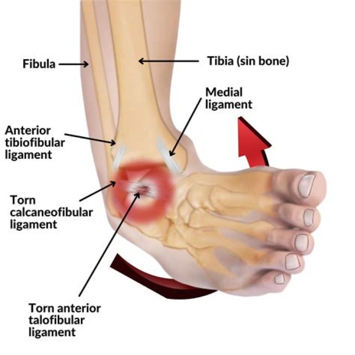
The calcaneofibular ligament crosses both the ankle joint and the subtalar joint. Injury to this ligament does not often occur in isolation, but is usually in conjunction with the anterior talofibular ligament. The calcaneofibular ligament resists varus stresses to the calcaneum in dorsiflexion.
Can you tear your Calcaneofibular ligament?
The ligament is most commonly damaged during inversion injuries to the ankle, and usually there is an associated injury to the anterior talofibular ligament (ATFL). The CFL is rarely torn in isolation and only a few case reports exist.
What is a Calcaneofibular ligament tear?
Calcaneofibular ligament injuries typically occur along with an anterior talofibular ligament injury within the scope of a lateral ankle sprain. The typical mechanism is excessive supination of the rearfoot on an externally rotated lower leg, leading to an inversion-internal-rotation type injury 1,2.What are the 3 bones that make up the ankle?
- The shin bone (tibia)
- The thinner bone running next to the shin bone (fibula)
- A foot bone that sits above the heel bone (talus)
The calcaneofibular ligament has its superior attachment on the malleolar fossa on the lateral malleolus, and its inferior attachment on the lateral surface of the calcaneus. Running across the calcaneofibular ligament are the tendons of the peroneus longus and peroneus brevis.
Article first time published onWhen should I get an MRI on my ankle?
Your doctor may recommend an MRI scan to help pinpoint the cause of your symptoms, particularly if they don’t improve after four to six weeks. It may be ordered to detect stress fractures in the foot or a cartilage or tendon injury, which can cause symptoms similar to those of a sprain.
How do I know if I have a Grade 3 ankle sprain?
Grade 3: This is a full tear of the ankle ligament. You may have heard a popping sound when it happened. This level of sprain causes severe pain, swelling and bruising. Because the ligament is no longer able to do its job, your ankle will feel unstable and will be unable to support any of your weight.
What does the talar tilt test for?
Purpose: To test for injury to the lateral ligaments of the ankle. Test Position: Supine or sitting. Performing the Test: The examiner stabilizes the distal leg in a neutral position and inverts the ankle. The examiner then determines how much inversion is present.
What does it mean when your talus hurts?
Osteochondral Lesions of the Talus (OLT) A sudden injury like a sprain can damage cartilage on your talus (heel bone) or cause fractures, blisters or sores in the bone underneath. You might notice a catch in your ankle, or it could lock up or still hurt months after a treated injury, which could be an OLT.
When is a talar tilt test positive?
The test is positive if, when compared with the opposite ankle, the talar tilt is 10°s or more. A 20° talar tilt indicates a positive test, regardless of comparison with the opposite ankle.
How do I know if my fiber is cotton?
Place a piece of the fabric in your fireproof container and ignite one corner. Pay attention to the odor of the smoke. Cotton smells like burning paper and has an afterglow at the end of the burn. An odor similar to burning hair or feathers indicates wool or silk fibers, but silk doesn’t always burn as easily as wool.
How do you measure tibiofibular clear space?
Measurement. The horizontal distance between the deepest point of the fibular groove or posterior tibial tubercle and the medial edge of the distal fibula 1 cm above the tibial plafond are measured in a normal anteroposterior view of the ankle 1-3.
How long does it take for syndesmosis to heal?
How long does syndesmosis take to heal? Syndesmosis injuries generally involve a period of immobilisation for 4-6 weeks depending on severity. (Yes that means a moon boot!) Following this we allow a further 6-12 weeks for a full recovery.
Which is the most common ligament to tear in ankle?
The most common and significant ligament tears include tears in the Anterior TaloFibular Ligament (ATFL), CalcaneoFibular Ligament (CFL), and the large Deltoid ligament complex.
Why do my ankles hurt after a long walk?
If the large ankle joints have arthritis, either at the tibiotalar or the subtalar joint, that can cause pain. There are also tendons on the inside and the outside of the ankle, and these can become inflamed and painful. By far, however, the most common cause of an ankle sore after walking is instability.
Where is the posterior Talofibular ligament?
Posterior talofibular ligament. The posterior talofibular ligament originates from the malleolar fossa, located on the medial surface of the lateral malleolus, coursing almost horizontally to insert in the posterolateral talus.
What is an eversion ankle sprain?
Eversion ankle sprains — occurs when the ankle rolls outward and tears the deltoid ligaments. Inversion ankle sprains — occurs when you twist your foot upward and the ankle rolls inward.
Can you use kinesiology tape for sprained ankle?
The use of kinesiology tape for the treatment of a lateral ankle sprain can be extremely effective.
What is foot eversion?
Foot eversion is when your foot collapses inward, usually with your feet also flattening. The sole of the foot actually faces away from your other foot, increasingly so as the problem worsens. … Many people think foot eversion is normal; it is not.
What is dorsiflexion and plantar flexion?
The term plantar flexion refers to the movement of the foot in a downward motion away from the body. … It also enables the opposite movement, dorsiflexion, which is the movement of the foot toward the leg. Your ankle joint supplies the power for 40% to 70% of your forward movement during walking.
What is dorsiflexion of the ankle?
Dorsiflexion is the backward bending and contracting of your hand or foot. This is the extension of your foot at the ankle and your hand at the wrist. … Dorsiflexion occurs in your ankle when you draw your toes back toward your shins. You contract the shinbones and flex the ankle joint when you dorsiflex your foot.
What is the lump on the side of my ankle called?
The most common fracture is to the bony bump on the outside of the ankle, the lateral malleolus. The lateral malleolus is the bottom of the fibula, the smaller lower leg bone. The bump on the inside of your ankle, the medial malleolus, is less commonly fractured.
What are the 7 bones in the ankle called?
The tarsal bones are 7 in number. They are named the calcaneus, talus, cuboid, navicular, and the medial, middle, and lateral cuneiforms.
Which bone is called the sit down bone?
ischium. sit down bone of the coxal bone.
What does Calcaneofibular ligament connect?
The calcaneofibular ligament (CFL), which connects the calcaneus, or heel bone, to the fibula.
What ligaments make up the Calcaneofibular ligament?
Calcaneofibular ligamentTofibula (lateral malleolus)IdentifiersLatinligamentum calcaneofibulareTA98A03.6.10.011
