In the adult central nervous system (CNS
Do ependymal cells form blood-brain barrier?
CSF functions as a cushion for the brain and spinal cord and provides important nutrients. … However, the tight junctions of the ependymal cells of the choroid plexuses form the blood–CSF barrier [180]. Ependymal cells of the choroid plexus are epithelial-like.
What are ependymal cells in the brain?
ependymal cell, type of neuronal support cell (neuroglia) that forms the epithelial lining of the ventricles (cavities) in the brain and the central canal of the spinal cord.
Which cells help form the blood-brain barrier?
Endothelial cells line the interior of all blood vessels. In the capillaries that form the blood–brain barrier, endothelial cells are wedged extremely close to each other, forming so-called tight junctions.Are ependymal cells part of the nervous system?
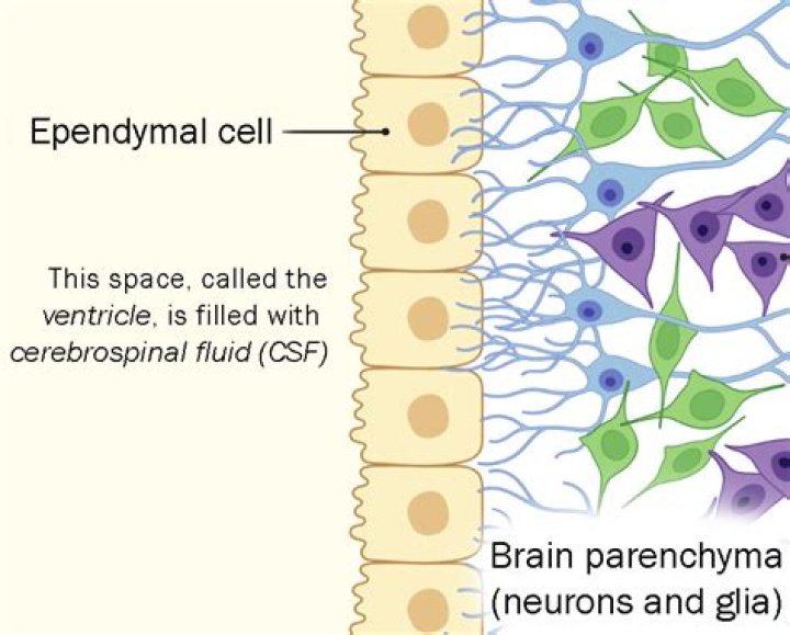
Ependymal cells are one of the four types of glial cells found in the central nervous system (CNS). Collectively, they form the ependyma which is a thin membrane that lines the cavities (or ventricles) in the brain and the central column of the spinal cord.
Where is the blood-brain barrier?
Where is the blood-brain barrier? The blood-brain barrier surrounds most of the blood vessels in the brain. It is a structure that is formed primarily due to the establishment of tight junctions between endothelial cells (i.e. cells that line the walls of blood vessels).
Is blood-brain barrier same as blood CSF barrier?
Blood–CSF Barrier Versus Blood–Brain Barrier: Fundamental Differences. Some authors consider the BCSFB as part of the generic BBB. As a first approximation, it is proved that both barriers have tight junctions (zonulae occludentes) that restrict the diffusion of most water-soluble solutes into the CNS.
What cells form the blood-brain barrier quizlet?
1. BBB endothelial cells, astrocytes, and neurons.Which cells maintain the blood-brain barrier quizlet?
Astrocytes, oligodendrocytes, microglia and ependymal cells. controlling interstitial environment, maintaining the blood brain barrier, creating three dimensional framework for CNS, performing repairs in damaged neural tissues and guides neuron development.
What does the blood-brain barrier separate?The blood-brain barrier is the barrier between the cerebral capillary blood and the interstitial fluid of the brain. … It functions as a semipermeable membrane that separates the peripheral blood from the cerebrospinal fluid (CSF) to maintains homeostasis within the central nervous system.
Article first time published onAre ependymal cells glia?
Ependymal cells are ciliated glial cells that form an epithelial barrier, called the ependyma, lining the brain’s ventricular system and the spinal cord’s central canal.
Are ependymal cells in the CNS or PNS?
Neuroglia in the CNS include astrocytes, microglial cells, ependymal cells and oligodendrocytes. Neuroglia in the PNS include Schwann cells and satellite cells.
Where is ependyma found?
The ependyma constitute a ciliated epithelium that derives from the neuroepithelium during development and is located at the interface between the brain parenchyma and ventricles in the central nervous system (CNS).
Which cells line the cavities of brain and spinal cord and form and move CSF?
Microglia are the smallest form of neuroglia and proliferate in response to injury. They increase in size as they phagocytose degraded matter. There are three different types of the last type of neuroglia, the ependyma. They line cavities of brain and spinal cord and maintain cerebrospinal fluid (CSF) flow.
What type of cells line the ventricles of the brain quizlet?
Ependymal cells (ependymocytes) are low columnar to cuboidal epithelial cells lining the ventricles of the brain and central canal of the spinal cord.
Do ependymal cells absorb CSF?
Cerebrospinal fluid Lining the CSF-filled ventricles, and spinal canal, the ependymal cells play an important role in the production and regulation of CSF. … Their apical surfaces are also covered with microvilli, which absorb CSF.
How are the blood brain barrier and blood CSF barrier maintained?
The barrier and secretory function of the choroid plexus epithelial cells are maintained by the expression of numerous transport systems allowing the directed transport of ions and nutrients into the CSF and the removal of toxic agents out of the CSF.
What makes up blood CSF barrier?
The endothelium, therefore, does not form a barrier to the movement of small molecules. Instead, the blood—CSF barrier at the choroid plexus is formed by the epithelial cells and the tight junctions that link them. The other part of the blood—CSF barrier is the arachnoid membrane, which envelops the brain.
Which of the following are layers within the blood brain barrier BBB?
Three barrier layers contribute to the separation of the blood and neural tissues: (1) a highly specialized endothelial cells (EC) layer comprising the blood-brain barrier (BBB) and partitioning the blood and brain interstitial fluid, (2) The blood-CSF barrier (BCSFB) with the choroid plexus epithelium which secretes …
Do all parts of the brain have the blood-brain barrier?
All areas of the brain do not have a blood-brain barrier. The structures located at strategic positions in the midline of the ventricular system and lack the BBB are collectively referred to as circumventricular organs (CVOs).
What is the blood-brain barrier give its components and function?
The blood-brain barrier (BBB) is the specialized system of brain microvascular endothelial cells (BMVEC) that shields the brain from toxic substances in the blood, supplies brain tissues with nutrients, and filters harmful compounds from the brain back to the bloodstream.
Do platelets cross the blood-brain barrier?
Platelets penetrate the blood-brain barrier (BBB) and accumulate in the lipid rafts-rich areas in the model of neuroinflammation.
Which of the following glial cells are a an integral part of the blood-brain barrier?
Schwann cells wrap closely around axons as the axons pass through the Schwann cell cytoplasm. Astrocytes are star-shaped glial cells that have numerous processes extending from them. Their foot processes are an important component of the blood-brain barrier.
Which of the following does not describe the blood-brain barrier?
The structure that does not contribute to the blood-brain barrier is c) Meninges. The meninges are three membranes that cover the brain and the spinal cord, and do not contribute to the blood-brain barrier.
Which of the following describes the blood-brain barrier?
Which of the following best describes the role of the blood-brain barrier? The blood-brain barrier is formed by tight junctions between capillary endothelial cells thus preventing the movement of solutes between cells. … Which area of the brain controls such basic functions as breathing, swallowing, and vomiting?
What do glia cells do?
Primarily, glial cells provide support and protection to the neurons (nerve cells), maintain homeostasis, cleaning up debris, and forming myelin. They essentially work to care for the neurons and the environment they are in.
What are the two types of glia cells?
There are three types of glial cells in the mature central nervous system: astrocytes, oligodendrocytes, and microglial cells (Figure 1.4A—C). Astrocytes, which are restricted to the brain and spinal cord, have elaborate local processes that give these cells a starlike appearance (hence the prefix “astro”).
What is the difference between Schwann cells and oligodendrocytes?
Oligodendrocytes are the cells which create myelin sheath around the axons of central nervous system. Schwann Cells are the cells which create myelin sheath around the axons of peripheral nervous system. Oligodendrocytes’ main function is the insulation of the nerve axons in the central nervous system.
Which of the following neuroglia is not part of the CNS?
Satellite cells are not a neuroglia of the CNS. Satellite cells support cell bodies of neurons in the peripheral nervous system. Astrocytes…
Are Schwann cells in the CNS or PNS?
Central nervous system (CNS) is composed of two major cell types: neuron and glia. … Consistently, the CNS houses oligodendrocytes for myelin synthesis. On the other hand, Schwann cells are the myelinating cells in the peripheral nervous system (PNS).
What is the name of the CNS neuroglia cell that acts as a phagocyte?
Astrocytes form a myelin sheath around axons in both the peripheral nervous system and the central nervous system. Schwann cells are the same as oligodendrocytes, and they both act as phagocytic cells to provide protection for the neuron.
